Harvard professor Jennifer Lewis has created a patch of tissue containing skin cells and biological structural material interwoven with blood-vessel-like structures using a 3-D printer and “disappearing” ink.
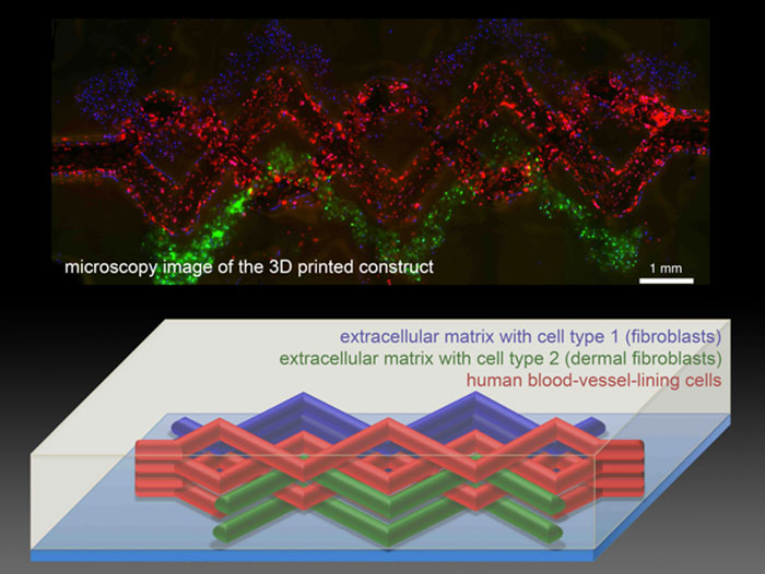
Lewis’s team created hollow, tube-like structures within a mesh of printed cells using an “ink” that liquefies as it cools. The tissue is built by the 3-D printer in layers. A gelatin-based ink acts as extracellular matrix—the structural mix of proteins and other biological molecules that surrounds cells in the body. Two other inks contained the gelatin material and either mouse or human skin cells. All these inks are viscous enough to maintain their structure after being laid down by the printer.
A third ink with counterintuitive behavior helped them create the hollow tubes. This ink has a Jell-O-like consistency at room temperature, but when cooled it liquefies. The team printed tracks of this ink amongst the others. After chilling the patch of printed tissue, the researchers applied a light vacuum to remove the special ink, leaving behind empty channels within the structure. Then cells that normally line blood vessels in the body can be infused into the channels.
The smallest channels printed were about 75 micrometers in diameter, which is much larger than the tiny capillaries that exchange nutrients and waste throughout the body. The hope is that the 3-D printing method will set the overall architecture of blood vessels within artificial tissue and then smaller blood vessels will develop along with the rest of the tissue.