Categories
Cancer Heart Sensors

Wearable sensor evaluates human tissue stiffness

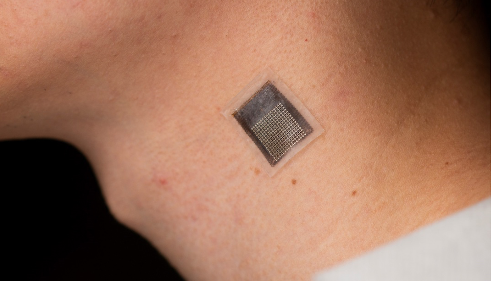
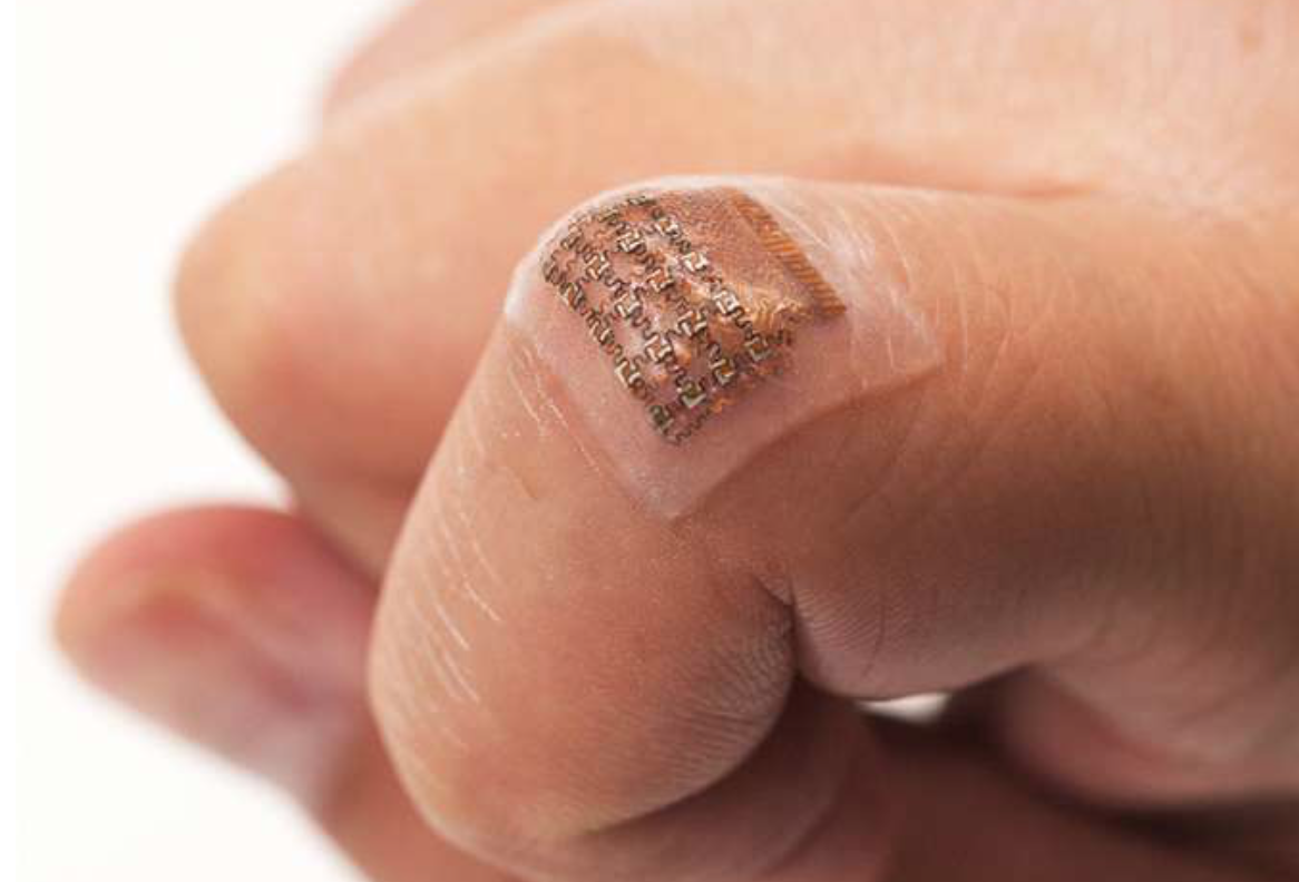
Sheng Xu and colleagues have developed a wearable, stretchable device that non-invasively evaluates the stiffness of human tissue, at an improved penetration depth, and for a longer period than, existing methods.

An ultrasonic array facilitates serial, non-invasive, three-dimensional imaging of tissues, four centimeters below the surface of human skin, at a spatial resolution of 0.5 millimeters.

The sensor can be used to detect cancer progression, which cases cells to stiffen; diagnose and treat sports injuries, by monitoring muscles, ligaments and tendons; and monitor the efficacy of treatments for liver, cardiovascular disease, and cancer, which cause tissue to stiffen.

Categories
Heart Sensors

Proof of concept wrist sensor could detect heart-vessel blockage in 3-5 minutes

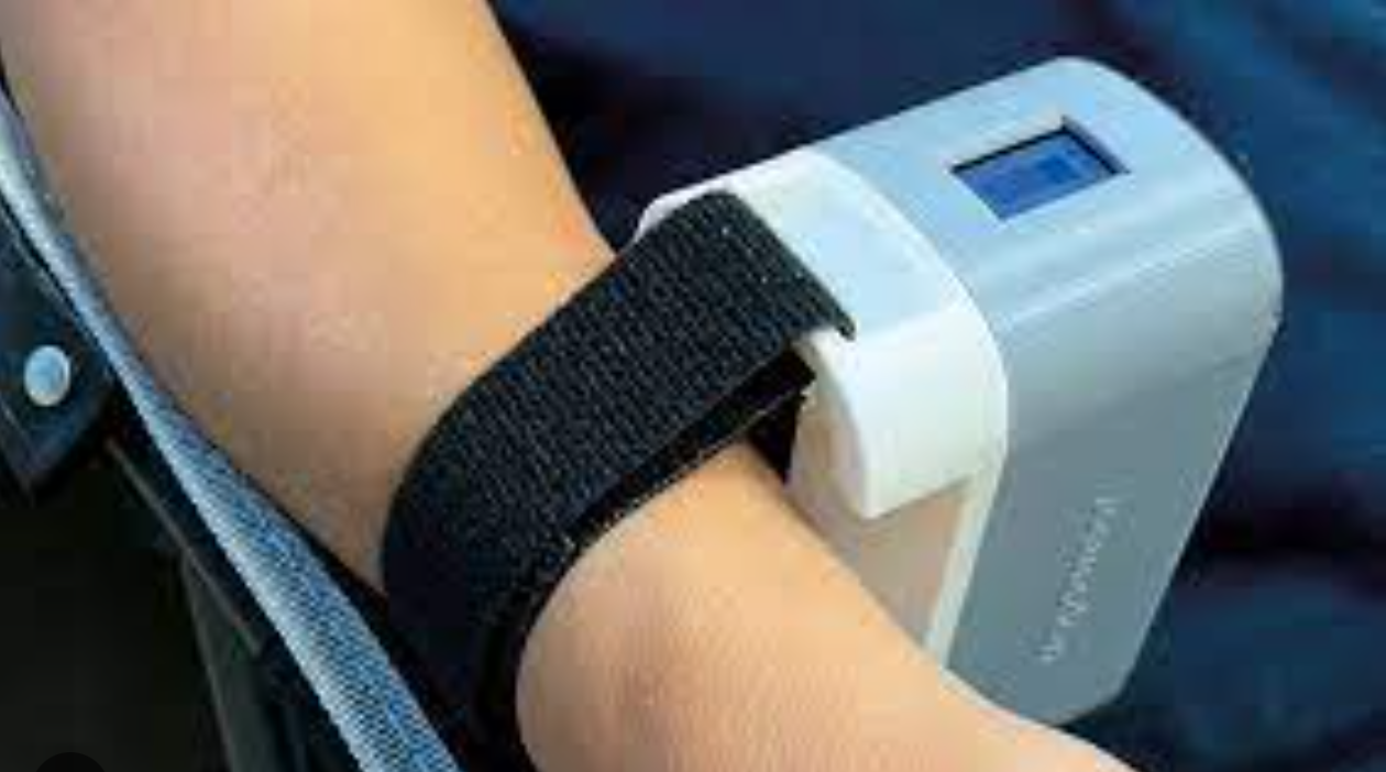
UW’s Graham Nichol and Harborview Medical Center colleagues studying the reliability of a troponin-detecting, wrist-worn sensor in arriving cardiac arrest patients.

Identifying heart-vessel blockage quickly is crucial to ensuring rapid, appropriate intervention. Traditional EKG diagnosis can lack accuracy, and blood testing for troponin can take hours.

The novel “Tropsensor,” being commercialized by rce, is designed to detect high troponin levels within three to five minutes of application, will be tested on 30 patients during emergency care at Harborview.

In a related Robert Wood Johnson study of 238 patients led by Partho Sengupta, a wrist worn transdermal infrared spectrophotometric sensor successfully identified elevated high-sensitivity cardiac troponin-I (hs-cTnI) levels in patients hospitalized with ACS.

Categories
Heart Ultrasound Wearables

Wearable cardiac ultrasound continuously monitors heart structure, function for 24 hours

UCSD professor Sheng Xu has developed a wearable ultrasound device that can continuously monitor and assess the structure and function of the human heart for 24 hours, during normal daily activity. This could eliminate the need for highly trained technicians and bulky devices. 

Signs of cardiac diseases are transient and unpredictable, and imaging can detect issues before they become problems. The new system gathers information through a small, soft, wearable patch, worn on the chest and designed for optimal adherence. It sends and receives ultrasound waves which are used to generate a constant stream of images of the structure of the heart in real time — with out radiation. As heart function issues often manifest only when the body is in motion, the wearable aspect of the device is particularly important.

Images of the four chambers of the heart are captured in different angles, and a clinically relevant subset of the images is analyzed. The model automatically segments the shape of the left ventricle from the image recording, extracting its volume frame-by-frame and yielding waveforms to measure stroke volume, cardiac output and ejection fraction. Curves of these three indices are generated continuously and noninvasively.

Xu plans to commercialize this technology through Softsonics, a company spun off from UC San Diego.


Sheng Xu was a featured speaker at the 2020 ApplySci Deep Tech Health conference on Sand Hill Road.

Categories
Heart Sensors

Wearable sensor network informs transient pacemaker

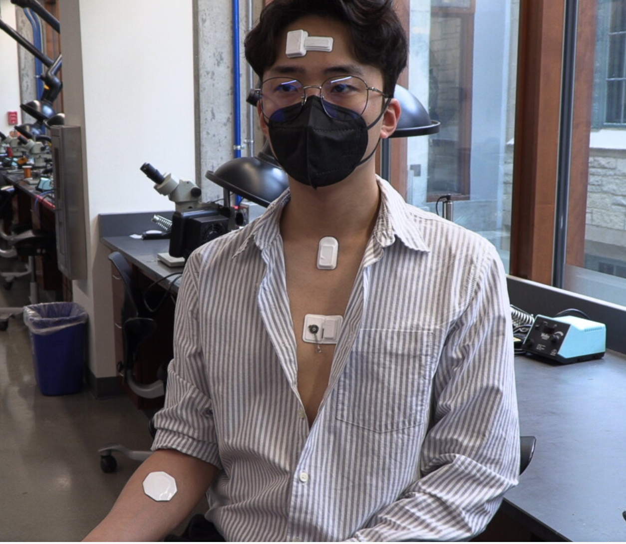
John Rogers and Northwestern colleagues have developed their second generation transient pacemaker, used post cardiac-surgery or for those awaiting permanent pacemakers. The new version, still implantable, wireless, and self dissolving, works with a network of soft wearable sensors, placed around the body.

The sensors continuously monitor body temperature, oxygen levels, respiration, muscle tone, physical activity, and cardiac electrical activity. Algorithms analyze the data to detect abnormal cardiac rhythms and decide when to pace the heart and at what rate. Physicians can remotely monitor the process through a phone or tablet. Energy is harvested, wirelessly, from a node within the network. Haptic feedback alerts wears of defects.

Rogers said: “This marks the first time we have paired soft, wearable electronics with transient electronic platforms. This approach could change the way patients receive care providing multimodal, closed-loop control over essential physiological processes — through a wireless network of sensors and stimulators that operates in a manner inspired by the complex, biological feedback loops that control behaviors in living organisms. For temporary cardiac pacing, the system untethers patients from monitoring and stimulation apparatuses that keep them confined to a hospital setting. Instead, patients could recover in the comfort of their own homes while maintaining the peace of mind that comes with being remotely monitored by their physicians. This also would reduce the cost of health care and free up hospital beds for other patients.”

The “body-area network” includes:

  • A battery-free transient, bioresorbable pacemaker to temporarily pace the heart
  • A cardiac module that sits on the chest to provide power to and control stimulation parameters for the implanted pacemaker as well as sense electrical activity and sounds of the heart
  • A hemodynamics module that sits on the forehead to sense pulse oximetry, tissue oxygenation and vascular tone
  • A respiratory module that sits at the base of the throat to monitor coughing and respiratory activity
  • A multi-haptic-feedback module that vibrates and pulses in a variety of patterns to communicate with the patient.

Rogers’ vision is of “multiple bioelectronic devices all talking to one another and performing different functions at different relevant anatomical locations” is a frontier area that he will continue to pursue.


Join ApplySci at MIT on September 30, 2022 for Deep Tech Health + Neurotech Boston

Categories
Heart

First all-digital clinical trial studies app-driven physical activity interventions

Stanford’s Euan Ashley has conducted an entirely digital clinical trial using the MyHeart Counts app, which is being used for patient recruitment, consent and interventions, and returns data to participants.

 1075 participants completed at least one intervention, and 493 completed the entire trial.  The higher than normal completion rate was attributed to the ease of enrollment and use.

The digital trial “prescribed” one of four simple interventions weekly, including  reminders to walk more or stand up. The team saw a 10% increase in activity compared to baseline.

The study serves as a template for future all-digital randomized clinical trials, which can include more nuanced questions, and an increase in interventions, which are ideally personalized.


Join ApplySci at the 12th Wearable Tech + Digital Health + Neurotech Boston conference on November 14, 2019 at Harvard Medical School featuring talks by Brad Ringeisen, DARPA – Joe Wang, UCSD – Carlos Pena, FDA  – George Church, Harvard – Diane Chan, MIT – Giovanni Traverso, Harvard | Brigham & Womens – Anupam Goel, UnitedHealthcare  – Nathan Intrator, Tel Aviv University | Neurosteer – Arto Nurmikko, Brown – Constance Lehman, Harvard | MGH – Mikael Eliasson, Roche – Nicola Neretti, Brown

Join ApplySci at the 13th Wearable Tech + Neurotech + Digital Health Silicon Valley conference on February 11-12, 2020 on Sand Hill Road featuring talks by Zhenan Bao, Stanford – Rudy Tanzi, Harvard – Shahin Farshchi – Lux Capital – Sheng Xu, UCSD – Carla Pugh, Stanford – Nathan Intrator, Tel Aviv University | Neurosteer – Wei Gao, Caltech

Categories
AI Heart

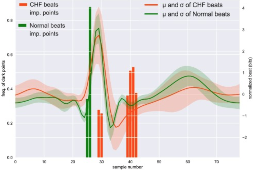
AI detects CHF through analysis of one heartbeat

Sebastiano Massaro at the University of Surrey,  Mihaela Porumb and Leandro Pecchia at the University of Warwick, and Ernesto Iadanza at the University of Florence have developed an advanced signal processing and machine learning method to identify congestive heart failure with 100% accuracy through analysis of one raw ECG heartbeat.

REGISTRATION RATES INCREASE SEPTEMBER 20 | Join ApplySci at the 12th Wearable Tech + Digital Health + Neurotech Boston conference on November 14, 2019 at Harvard Medical School featuring talks by Brad Ringeisen, DARPA – Joe Wang, UCSD – Carlos Pena, FDA  – George Church, Harvard – Diane Chan, MIT – Giovanni Traverso, Harvard | Brigham & Womens – Anupam Goel, UnitedHealthcare  – Nathan Intrator, Tel Aviv University | Neurosteer – Arto Nurmikko, Brown – Constance Lehman, Harvard | MGH – Mikael Eliasson, Roche
Categories
Heart

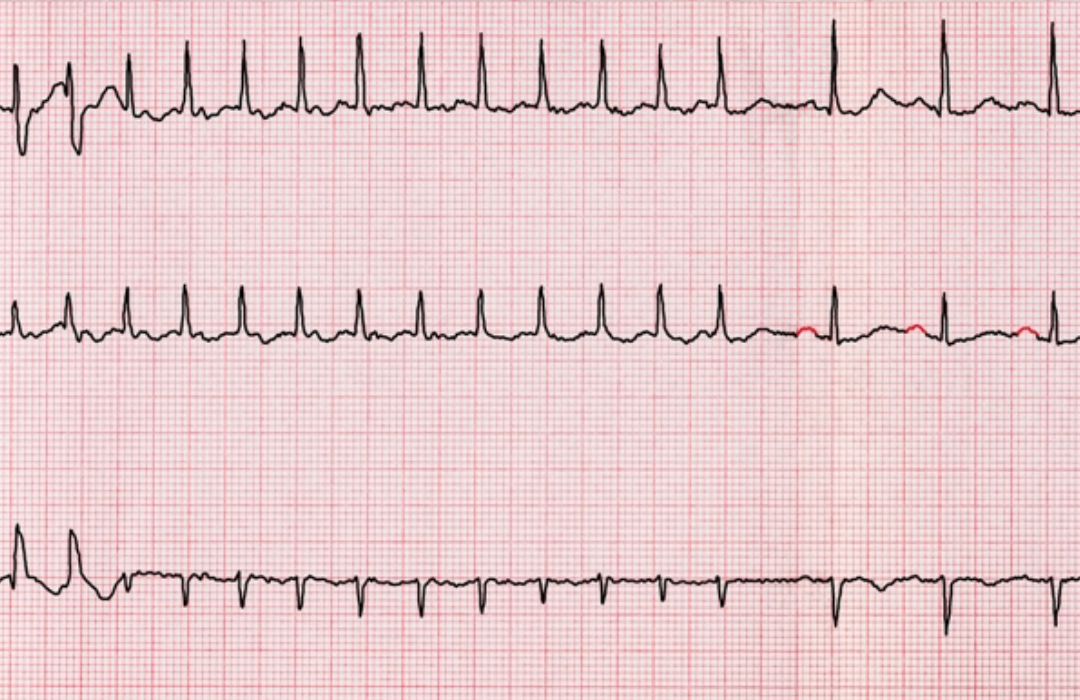
Atrial fibrillation-detecting ring

Eue-Keun Choi and Seoul National University colleages have developed an atrial fibrillation detecting ring, with similar functionality to AliveCor and other watch-based monitors. The researchers claim that the performance is comparable to medical grade pulse oximeters.

In a study, Soonil Kwon and colleagues analyzed data from 119 patients with AF who underwent simultaneous ECG and photoplethysmography before and after direct-current cardioversion. 27,569 photoplethysmography samples were analyzed by an algorithm developed with a convolutional neural network. Rhythms were then interpreted with the wearable ring.

The accuracy of the convolutional neural network was 99.3% to diagnose AF and 95.9% to diagnose sinus rhythm.  The accuracy of the wearable device was 98.3% for sinus rhythm and 100% for AF after filtering low-quality samples.

Choi believes that: “Deep learning or [artificial intelligence] can overcome formerly important problems of [photoplethysmography]-based arrhythmia diagnosis. It not only improves diagnostic accuracy in great degrees, but also suggests a metric how this diagnosis will be likely true without ECG validation. Combined with wearable technology, this will considerably boost the effectiveness of AF detection.”.


Join ApplySci at the 12th Wearable Tech + Digital Health + Neurotech Boston conference on November 14, 2019 at Harvard Medical School and the 13th Wearable Tech + Neurotech + Digital Health Silicon Valley conference on February 11-12, 2020 at Stanford University

Categories
3-D Printing Heart

3D printed, vascularized heart, using patient’s cell, biological materials

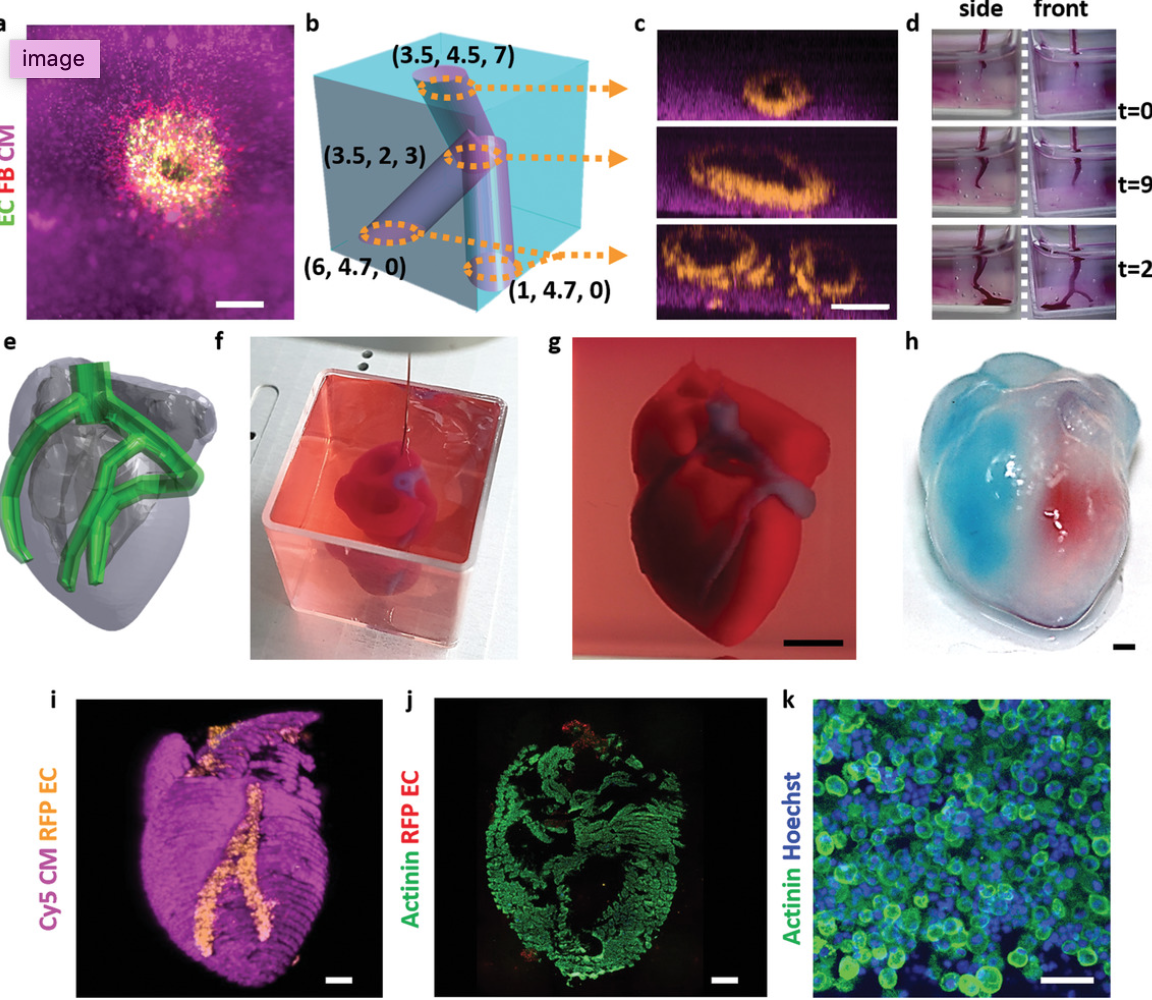
Tel Aviv University professor Tal Dvir has printed a 3D vascularized engineered heart, including cells, blood vessels, ventricles and chambers,  using a patient’s own cell and biological materials.

A biopsy of fatty tissue was taken from patients. Cellular and a-cellular materials were separated. While the cells were reprogrammed to become pluripotent stem cells, the extracellular matrix were processed into a personalized hydrogel that served as printing “ink.” After being mixed with the hydrogel, the cells were efficiently differentiated to cardiac or endothelial cells to create patient-specific, immune-compatible cardiac patches with blood vessels and, subsequently, an entire heart.

Dvir believes that this “3D-printed thick, vascularized and perfusable cardiac tissues that completely match the immunological, cellular, biochemical and anatomical properties of the patient” reduces the risk of implant rejection.

The team now plans on culturing the printed hearts and “teaching them to behave” like hearts, then transplanting them in animal models.


Join ApplySci at the 12th Wearable Tech + Digital Health + Neurotech Boston conference on November 14, 2019 at Harvard Medical School and the 13th Wearable Tech + Neurotech + Digital Health Silicon Valley conference on February 11-12, 2020 at Stanford University

Categories
Heart Sensors

Adhesive emergency response sensors

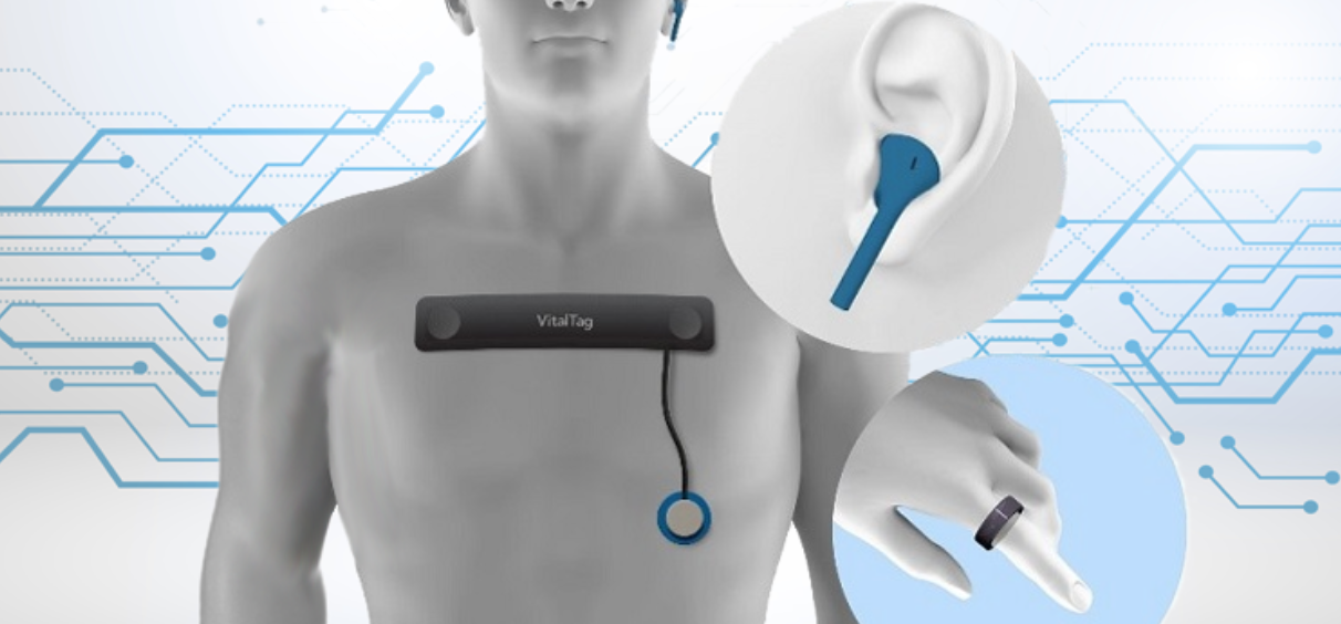
VitalTag by Pacific Northwest National Laboratory is a chest-worn sticker that detects, monitors and transmits blood pressure, heart rate, respiration rate and other vital signs, eliminating the need for multiple medical devices.

It is meant for emergency responders to quickly assess a person’s state.

Additional sensors are worn on the finger, and in the ear.

Data is displayed in an app, allowing responders to see patients’ location and receive alerts when status changes or they are moved.  Multiple patients can be monitored simultaneously.


Join ApplySci at the 9th Wearable Tech + Digital Health + Neurotech Boston conference on September 24, 2018 at the MIT Media Lab.  Speakers include:  Rudy Tanzi – Mary Lou Jepsen – George ChurchRoz PicardNathan IntratorKeith JohnsonJohn MattisonRoozbeh GhaffariPoppy Crum – Phillip Alvelda Marom Bikson – Ed Simcox – Sean Lane

Categories
Blood Pressure Heart Sensors

Small ultrasound patch detects heart disease early

Sheng Xu, Brady Huang, and UCSD colleagues have developed a small, wearable ultrasound patch that  monitors blood pressure in arteries up to 4 centimeters under the skin.  It is meant to detect cardiovascular problems earlier, with greater accuracy

Applications include continuous blood pressure monitoring in heart and lung disease, the critically ill, and those undergoing surgery.  It could be used to measure other vital signs, but this was not studied.

The wearable measures central blood pressure, considered more accurate and better at predicting disease than peripheral blood pressure. Central blood pressure is not routinely measured, and involves a catheter inserted into a blood vessel in the arm, groin or neck, and guiding to the heart. A non-invasive method exists, but it does not produce consistently accurate readings.


Join ApplySci at the 9th Wearable Tech + Digital Health + Neurotech Boston conference on September 24, 2018 at the MIT Media Lab.  Speakers include:  Rudy Tanzi – Mary Lou Jepsen – George ChurchRoz PicardNathan IntratorKeith JohnsonJohn MattisonRoozbeh GhaffariPoppy Crum – Phillip Alvelda Marom Bikson – Ed Simcox – Sean Lane

Categories
Heart Seniors

Apple watch detects falls, diagnoses heart rhythm, bp irregularities

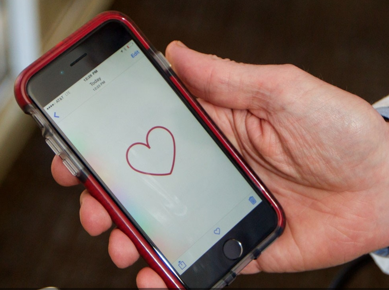
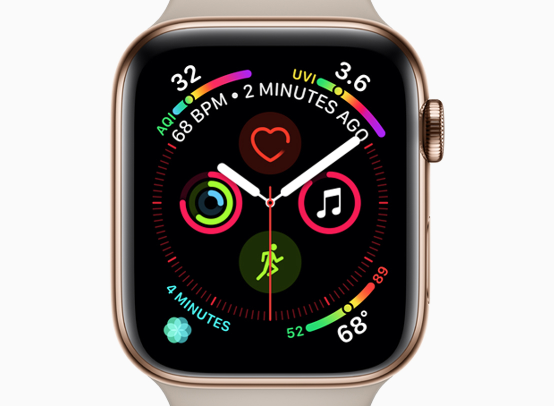
The Apple Watch has become a serious medical monitor.  It will now be able to detect falls, contact emergency responders, and diagnose  irregularities in heart rhythm and blood pressure.  Its ECG app has been granted a De Novo classification by the FDA.

ECG readings are taken from the wrist, using electrodes built into the Digital Crown and an electrical heart rate sensor in the back crystal. Users touch the Digital Crown and receive a heart rhythm classification in 30 seconds. It can classify if the heart is beating in a normal pattern or whether there are signs of Atrial Fibrillation . All recordings, their associated classifications and any noted symptoms are stored and can be shared with physicians.

The watch intermittently analyzes heart rhythms in the background and sends a notification if an irregular heart rhythm such as AFib is detected.  It can also alert the user if the heart rate exceeds or falls below a specified threshold.

Fall detection is via a built in accelerometer and gyroscope, which measures forces, and an algorithm to identify hard falls. Wrist trajectory and impact acceleration are analyzed to detect falls.  Users are then sent an alert, which can be dismissed or used to call emergency services.  If  immobility  is sensed for 60 seconds,  emergency services will automatically be called, and emergency contacts will be notified.


Join ApplySci at the 9th Wearable Tech + Digital Health + Neurotech Boston conference on September 24, 2018 at the MIT Media Lab.  Speakers include:  Rudy Tanzi – Mary Lou Jepsen – George ChurchRoz PicardNathan IntratorKeith JohnsonJohn MattisonRoozbeh GhaffariPoppy Crum – Phillip Alvelda Marom Bikson – Ed Simcox – Sean Lane
Categories
Brain Heart Virtual Reality

David Axelrod: VR in healthcare & the Stanford Virtual Heart | ApplySci @ Stanford

David Axelrod discussed VR-based learning in healthcare, and the Stanford Virtual Heart, at ApplySci’s recent Wearable Tech + Digital Health + Neurotech conference at Stanford;


Join ApplySci at the 9th Wearable Tech + Digital Health + Neurotech Boston conference on September 24, 2018 at the MIT Media Lab.  Speakers include:  Rudy Tanzi – Mary Lou Jepsen – George ChurchRoz PicardNathan IntratorKeith JohnsonJuan EnriquezJohn MattisonRoozbeh GhaffariPoppy Crum – Phillip Alvelda Marom Bikson

REGISTRATION RATES INCREASE JULY 6th