Categories
AI Covid-19

AI detects COVID in chest x rays

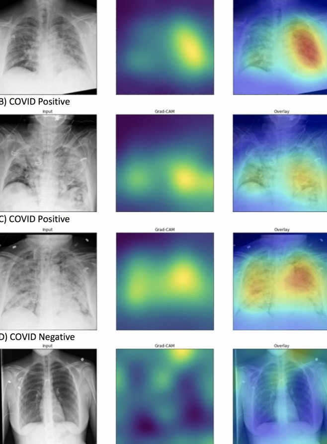
DeepCOVID-XR is a Northwestern University developed algorithm that automatically detects the signs of COVID-19 on a basic X-ray of the lungs. The system is able to detect COVID-19 in X-rays 10 times faster than thoracic radiologists and 1% to 6% more accurately.

The developers said the AI could be used to rapidly screen patients at hospital admission, and trigger protocols to help protect healthcare workers.

Accoring to Professor Aggelos Katsaggelos, the alorithm will not replace testing, but will enable the use of cheap, routine, safe x-rays to determine if a patient needs to be isolated.

In 17,000 X-ray images, the algorithm identified lungs which appeared patchy and hazy, as air sacs became inflamed and filled with fluid instead of air, which is common in COVID-19.

When put up against five experienced, fellowship-trained radiologists, DeepCOVID-XR was able to process a set of 300 test X-rays in about 18 minutes, compared to about two and a half to three and a half hours. The AI also delivered an accuracy rate of 82%, about on par with the group’s range of 76% to 81%.

“Radiologists are expensive and not always available,” Katsaggelos said. “X-rays are inexpensive and already a common element of routine care. This could potentially save money and time—especially because timing is so critical when working with COVID-19.”
Categories
Covid-19 Sensors

Electrostatic and electrochemical sensors rapidly detect airborne viruses

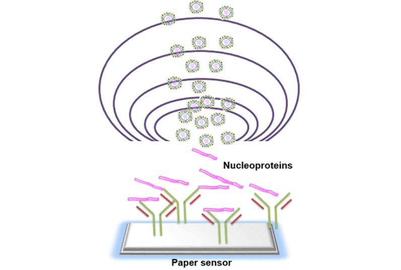
Jang Jae-sung and Ulsan colleagues have developed a method to quickly and accurately detect airborne viruses, to inform public health and quarantine efforts.

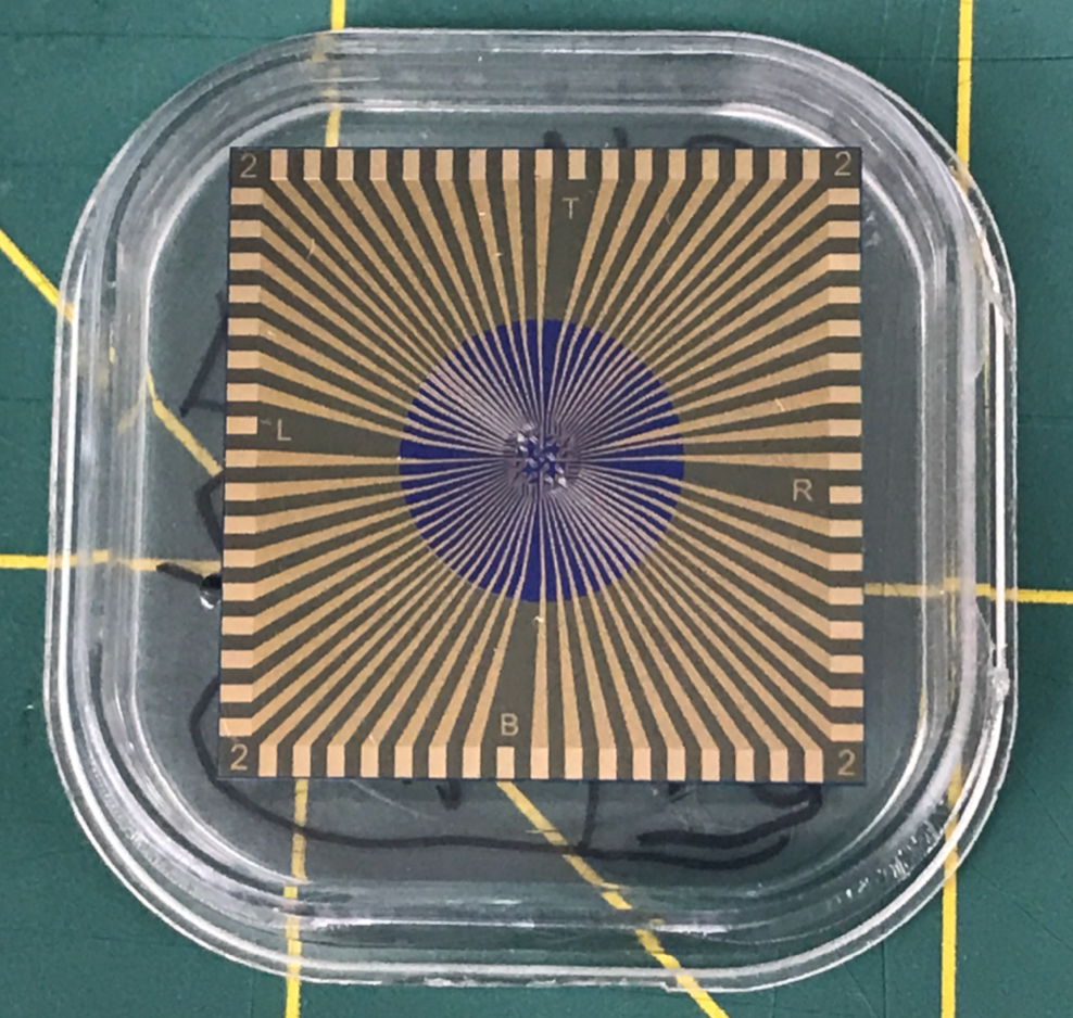
Electrostatic force captures and condenses viruses in the air, and a paper electrochemical sensor checks samples for antigens and virus antibodies. Liquid particles as small as 1 micrometer have been successfully collected.

Current methods of airborne sample collection typically use vacuums that can damage the samples, and cannot collect very small particles.

A recent study showed that tests on the type-A H1N1 flu virus showed good results. Jang believes that since the coronavirus is similar in structure and size, the technology should be applicable to COVID-19, which he is researching.

Categories
Covid-19 Sensors Wearables

Presymptomatic COVID detection with wearables

Stanford’s Michael Snyder and colleagues have used smartwatch data to detect early, presymptomatic COVID-19 in 31 individuals out of a cohort of 5,000.

They demonstrated that COVID-19 infections are associated with alterations in heart rate, steps and sleep in 80% of cases. Physiological alterations were detected prior to, or at, symptom onset in 85% of the positive cases, in some cases nine or more days before symptoms.

A method to detect onset of COVID-19 infection in real-time was developed, which detected 67% of infection cases at or before symptom onset.

The study intends to provide a roadmap to a rapid and universal diagnostic method for the large-scale detection of respiratory viral infections in advance of symptoms.

—————————————————————————-

Click to view Professor Snyder’s talk at the 2019 ApplySci conference at Stanford.

Categories
Covid-19 Sensors

First nutrition monitoring wearable tracks vitamin C; could be useful in COVID treatment

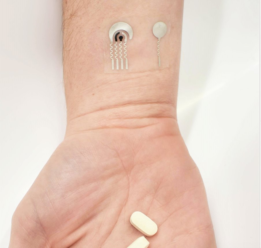
UCSD’s Joe Wang has again disrupted chemical sensing, by creating a wearable vitamin C sensor, which is a departure from now common vital sign and activity sensing wearables. This is the first time a wearable has been used to track nutritional intake, a key component of general health and disease prevention.

Vitamin C cannot be synthesized by the human body and must be obtained through food or supplements. It supports immune health, collagen production, wound healing and may be useful in treating cancer, heart disease, and COVID-19. High doses have been linked to reduced mortality in COVID patients with Sepsis and/or ARDS in studies.

The adhesive patch, applied to skin, stimulates sweating, and quickly detects vitamin c levels using flexible electrodes containing the enzyme ascorbate oxidase. When vitamin C is present, the enzyme converts it to dehydroascrobic acid. The resulting consumption of oxygen generates a current that is measured by the device. 

Click to view Prof Wang’s (brilliant) talk at the recent ApplySci conference at Harvard Medical School.

Categories
Covid-19 Sensors

Single sensor could simultaneously detect, differentiate between flu, coronavirus

University of Texas professor Deji Akinwande is developing a graphene sensor that can tell the difference between flu and coronavirus, and test for both simultaneously. The goal is to save time, medical resources, and cost, and speed appropriate treatment, as a second COVID wave could correspond with the next flu season.

The sensor is the size of a micro USB drive and is infused with antibodies of both COVID-19 and influenza. One part of the device is sensitive only to the flu, while another part will react only to the coronavirus.

The researchers will use inactive samples of COVID-19 and influenza for initial testing, and will measure how he sensor connects with the coronavirus’s spike proteins, which help it enter human cells by binding with them.

The work builds on the team‘s previous iron deficiency detecting graphene sensor work. “It became clear that just by changing the antibody, we could pivot the platform to focus on the coronavirus,” Akinwande said.

Categories
3-D Printing Covid-19 Sensors

Sensors 3D printed directly on lungs, heart could be used with surgical robots to diagnose, monitor disease

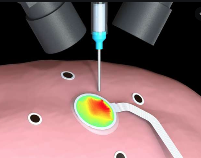
Michael McAlpine and University of Minnesota colleagues used 3D printing and motion capture technology to print electronic sensors directly on organs that are expanding and contracting, such as the heart and lungs. This could be used to diagnose and monitor the lungs of patients with COVID-19.

This builds on the team’s technique which enabled the printing of electronics directly on the skin of a hand that moved left to right or rotated.

They used a balloon-like surface and a specialized 3D printer, with motion capture tracking markers to help the 3D printer adapt its printing path to the expansion and contraction movements on the surface. An animal lung in the lab was artificially inflated and a soft hydrogel-based sensor was printed directly on the surface.

According to McAlpine, “the broader idea behind this research, is that this is a big step forward to the goal of combining 3D printing technology with surgical robots. In the future, 3D printing will not be just about printing but instead be part of a larger autonomous robotic system. This could be important for diseases like COVID-19 where health care providers are at risk when treating patients.”

Categories
Covid-19 Sensors

Organic electronic patch + algorithm continuously monitor multiple COVID symptoms

Northwestern and University of Illinois researchers have partnered to combine a COVID symptom-detecting wearable with a method to organize and analyze the massive data sets required to accurately show disease progression. The technology will be used in hospitals and nursing homes, to monitor both patients and healthcare workers, to identify contagion early in an effort to reduce the risk of spread.

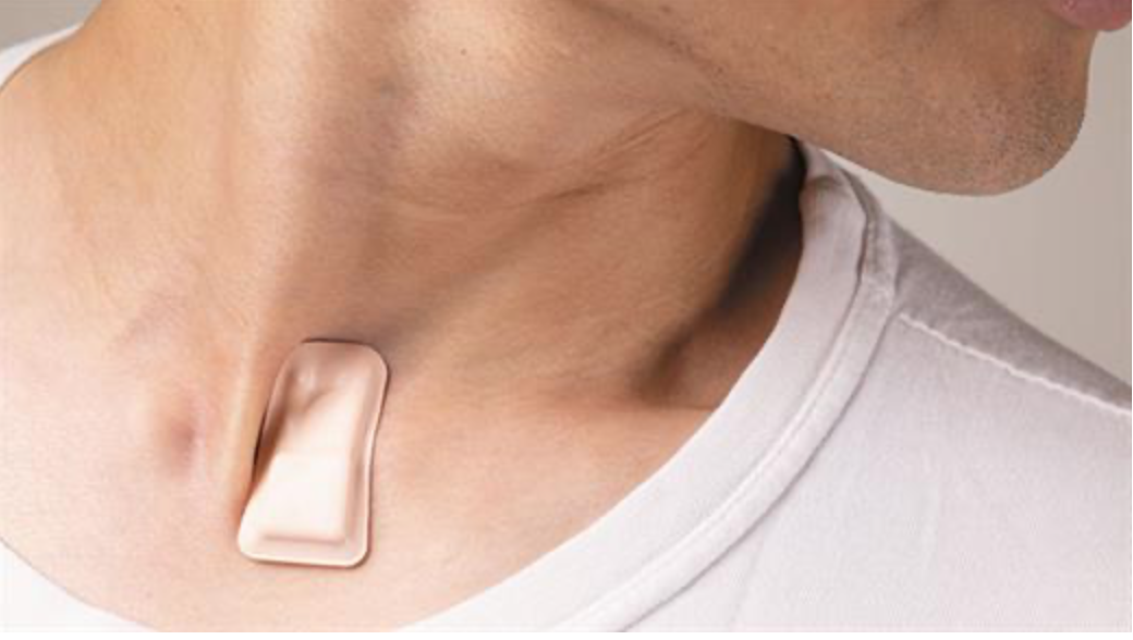
Building on his stroke-monitoring wearable, John Rogers at Northwestern uses a patch that sits at the base of the throat and continuously monitors cough, heart and respiratory sounds. The University of Illionois algorithm, developed by Naresh Shanbhag, will allow these parameters to be tracked quantitatively.

The goal is to extract detailed, and sometimes subtle, parameters from the raw data, which provide insights into disease progression. This includes analyzing cough intensity, whether it is wet or dry, and whether a patient swallows afterward,

In addition to hospital patients, the team was able to monitor a nurse before, during, and after she had contracted the virus. The data collected, in combination with the nurse’s detailed notes throughout her illness, allowed the team to pick up heart rate spikes and changes in coughing activity that would have gone undetected in standard non-ICU hospital care.

Data is transmitted from the patch to a phone or a tablet, and then to the cloud, where it is processed. The group is attempting to move the processing directly to the patch and device to save energy consumption, and improve security and privacy.

According to Shanbhag: “We will acquire data for both ill and healthy patients, and learn the characteristics of the data by developing COVID-19 specific, low-complexity machine learning algorithms. We’ll then use the learned models for predicting whether a patient is ill or not and how the disease will progress over time for new patients or individuals.”

Categories
Covid-19 Stem Cells

Stem cell treatment for ARDS in COVID-19 patients

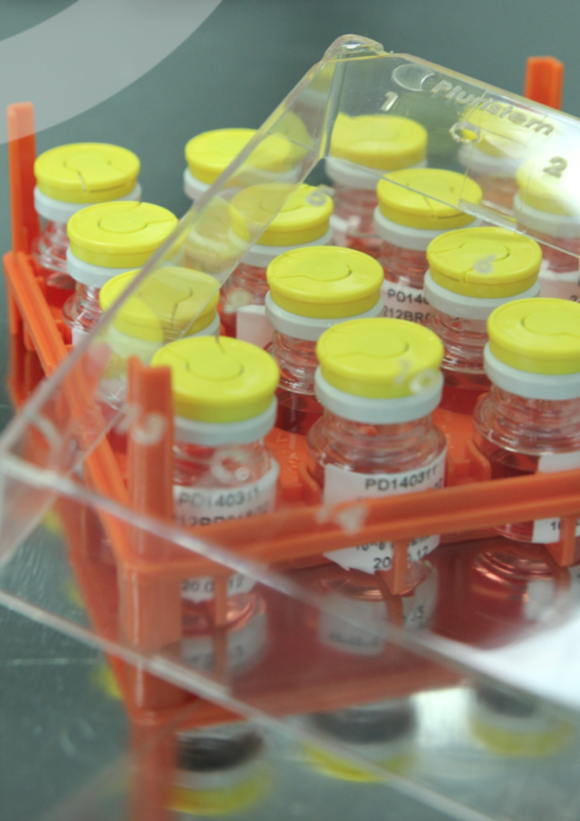
Last week, Pluristem released initial results from its compassionate use program for the treatment of patients with acute respiratory failure and inflammatory complications resulting from COVID-19. The treatment was administered in an Israeli hospital. All seven ICU patients with ARDS treated with Pluristem’s PLX cell therapy have survived. 6 out of 7 have completed the seven-day follow-up (one is still within the period. 4 out of the 6 (66%) patients displayed improvement in respiratory parameters, and 3 of the 6 (50%) patients are in late stages of weaning from ventilators.

Pluristem has now treated its first COVID-19 patient in the United States. under the FDA’s Single Patient Expanded Access Program, also called a compassionate use program, which is part of the U.S. Coronavirus Treatment Acceleration Program (CTAP.) The patient was treated with PLX cell therapy at Holy Name Medical Center in New Jersey, an acute care facility that is currently an active site for Pluristem’s Phase III critical limb ischemia (CLI) study. Prior to treatment with PLX, the patient was critically ill with respiratory failure due to acute respiratory distress syndrome (ARDS) and was under mechanical ventilation in an intensive care unit (ICU) for three weeks.

PLX cells are available off-the-shelf and once commercialized, can be manufactured in large scale quantities, offering an advantage in addressing a global pandemic. PLX cells are allogeneic mesenchymal-like cells that have immunomodulatory properties that induce the immune system’s natural regulatory T cells and M2 macrophages, and thus may prevent or reverse the dangerous overactivation of the immune system. PLX cells may reduce the incidence and\or severity of COVID-19 pneumonia and pneumonitis leading hopefully to a better prognosis for the patients. Previous pre-clinical findings of PLX cells revealed therapeutic benefit in animal studies of pulmonary hypertension, lung fibrosis, acute kidney injury and gastrointestinal injury which are potential complications of the severe COVID-19 infection. Clinical data using PLX cells demonstrated the strong immunomodulatory potency of PLX cells in patients post major surgery. This is a potential therapy for mitigating the tissue-damaging effects of COVID-19.

Categories
Covid-19

Continuous COVID-19 PPG vital sign monitoring in hospital and home

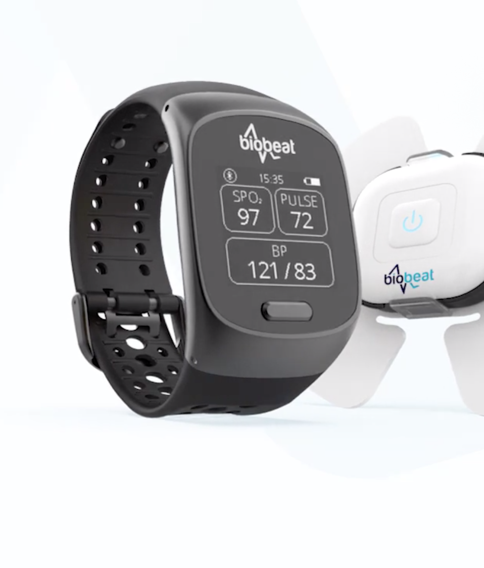
Biobeat‘s wrist wearable uses PPG wave reading for continuous, cuffless, noninvasive medical-grade monitoring of blood pressure, oxygen saturation, respiratory rate, heart rate, temperature and other vitals. It is being widely used for Israeli COVID-19 patients in hospitals and at home.

Categories
Covid-19

UCSF/Oura ring COVID-19 onset, progression, recovery study

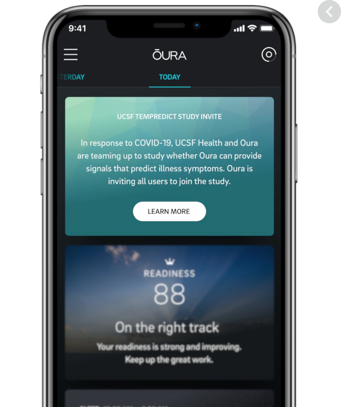
UCSF’s Ashley Mason is using the Oura Ring to build an algorithm to help identify patterns of onset, progression, and recovery, for COVID-19.

The study will combine physiological data (temperature, respiratory rate, heart rate) with responses to daily symptom surveys from 2,000 front-line healthcare workers and the general population. It will be open to all Oura ring users.

The approach, if successful, could be used to track and manage other illnesses and conditions.

Click for details of the TemPredict study.