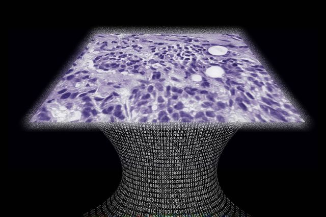
UCLA professor Aydogan Ozcan has developed a lens-free microscope for high throughput 3-D tissue imaging to detect cancer or other cell level abnormalities.
Laser or light-emitting-diodes illuminate a tissue or blood sample on a slide inserted into the device. A sensor array on a microchip captures and records the pattern of shadows created by the sample. The patterns are processed as a series of holograms, forming 3-D images of the specimen. An algorithm color codes the reconstructed images. Contrasts in the samples more apparent than they would be in the holograms, detecting abnormalities.
This could lead to cheaper and more portable technology for examining tissue, blood and other biomedical specimens. It will benefit patients in remote areas and in cases where large numbers of samples need to be examined quickly.
