https://www.einstein.yu.edu/news/releases/968/watching-molecules-morph-into-memories/
Albert Einstein College of Medicine researchers have used advanced imaging techniques to observe memory making molecules travel in real time in living brain cells.
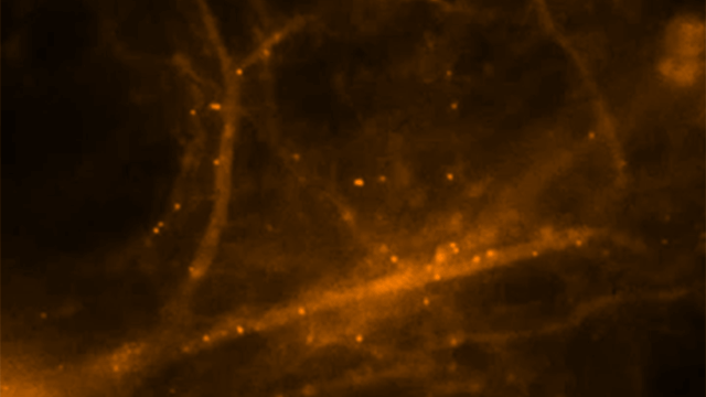
To look deeply into neurons without harming them, the researchers developed a mouse model in which they fluorescently tagged all molecules of messenger RNA that code for beta-actin protein – an essential structural protein found in large amounts in brain neurons and considered a key player in making memories. mRNA is a family of RNA molecules that copy DNA’s genetic information and translate it into the proteins that make life possible. They stimulated neurons from the mouse’s hippocampus, where memories are made and stored, and then watched fluorescently glowing beta-actin mRNA molecules form in the nuclei of neurons and travel within dendrites, the neuron’s branched projections. They discovered that mRNA in neurons is regulated through a novel process described as “masking” and “unmasking,” which allows beta-actin protein to be synthesized at specific times and places and in specific amounts.
Neurons come together at synapses, where slender dendritic “spines” of neurons grasp each other, much as the fingers of one hand bind those of the other. Evidence indicates that repeated neural stimulation increases the strength of synaptic connections by changing the shape of these interlocking dendrite “fingers.” Beta-actin protein appears to strengthen these synaptic connections by altering the shape of dendritic spines. Memories are thought to be encoded when stable, long-lasting synaptic connections form between neurons in contact with each other.
Hye Yoon Park stimulated individual hippocampal neurons of the mouse and observed newly formed beta-actin mRNA molecules within 10 to 15 minutes, indicating that nerve stimulation had caused rapid transcription of the beta-actin gene. Further observations suggested that these beta-actin mRNA molecules continuously assemble and disassemble into large and small particles, respectively. These mRNA particles were seen traveling to their destinations in dendrites where beta-actin protein would be synthesized.
Adina Buxbaum showed that neurons may be unique among cells in how they control the synthesis of beta-actin protein. This revealed the mechanism by which brain neurons handle this challenge. She found that as soon as beta-actin mRNA molecules form in the nucleus of hippocampal neurons and travel out to the cytoplasm, the mRNAs are packaged into granules and so become inaccessible for making protein. She then saw that stimulating the neuron caused these granules to fall apart, so that mRNA molecules became unmasked and available for synthesizing beta-actin protein.
Leave a Reply