Categories
AI Brain Cancer

AI decodes brain tumor DNA during surgery

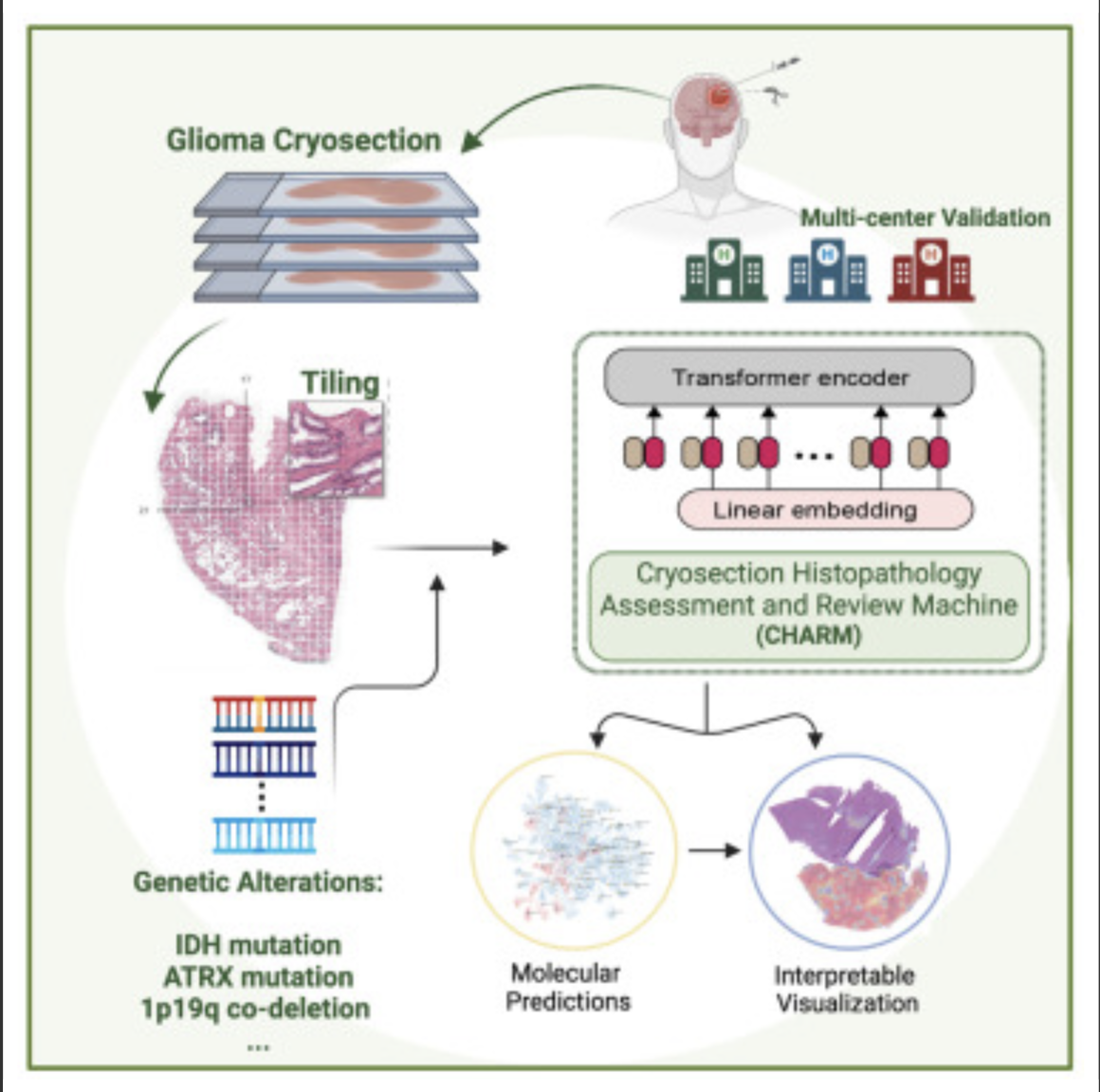
Kun-Hsing Yu and HMS colleagues used AI to rapidly determine a brain tumor’s molecular identity during surgery, propeling the development of precision oncology. The tool is CHARM (Cryosection Histopathology Assessment and Review Machine.) Currently, genetic sequencing takes days to weeks.  

Accurate molecular diagnosis during surgery can help a neurosurgeon decide how much brain tissue to remove. Removing too much when the tumor is less aggressive can affect a patient’s neurologic and cognitive function. Removing too little when the tumor is highly aggressive may leave behind malignant tissue that can grow and spread quickly. 

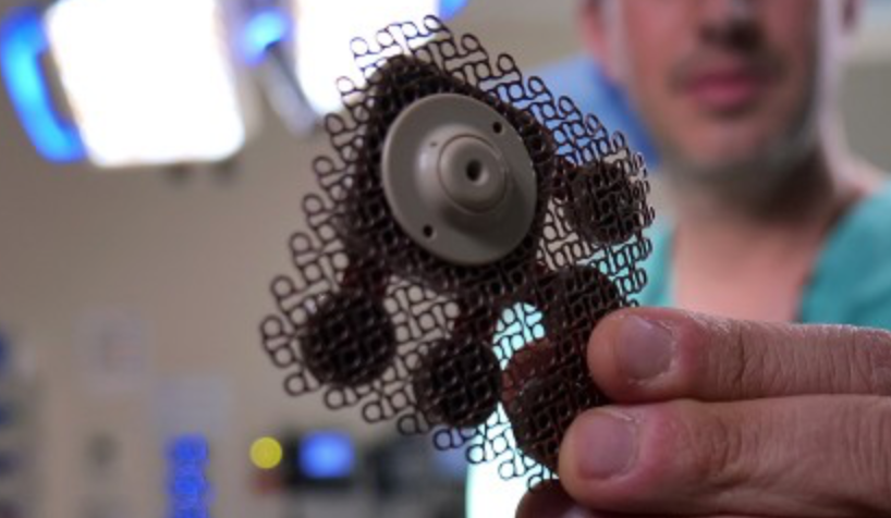
The technology will also allow the surgeon to determine if the patient can benefit from immediate treatment with drug-coated wafers placed directly into the brain at the time of the operation.

The standard intraoperative diagnostic approach used now involves taking brain tissue, freezing it, and examining it under a microscope. A major drawback is that freezing the tissue tends to alter the appearance of cells under a microscope and can interfere with the accuracy of clinical evaluation. Furthermore, the human eye, even when using potent microscopes, cannot reliably detect subtle genomic variations on a slide.

The new AI approach overcomes these challenges and could be particularly valuable in areas with limited access to technology to perform rapid cancer genetic sequencing.

Knowledge of a tumor’s molecular type provides insight about its aggressiveness, behavior, and likely response to various treatments, which can inform post-operative decisions.

The new tool enables during-surgery diagnoses aligned with the WHO classification system for diagnosing and grading the severity of gliomas, which calls for such diagnoses to be made based on a tumor’s genomic profile.

CHARM was developed using 2,334 brain tumor samples from 1,524 people with glioma from three different patient populations. When tested on a never-before-seen set of brain samples, the tool distinguished tumors with specific molecular mutations at 93 percent accuracy and successfully classified three major types of gliomas with distinct molecular features that carry different prognoses and respond differently to treatments.

It successfully captured visual characteristics of the tissue surrounding the malignant cells. It was capable of spotting telltale areas with greater cellular density and more cell death within samples, both of which signal more aggressive glioma types.

CHARM was also able to pinpoint clinically important molecular alterations in a subset of low-grade gliomas, a subtype of glioma that is less aggressive and therefore less likely to invade surrounding tissue. Each of these changes also signals different propensity for growth, spread, and treatment response.

It further connected the appearance of the cells — the shape of their nuclei, the presence of edema around the cells — with the molecular profile of the tumor. This means that the algorithm can pinpoint how a cell’s appearance relates to the molecular type of a tumor.

Accorging to Yu, this ability to assess the broader context around the image renders the model more accurate and closer to how a human pathologist would visually assess a tumor sample.

The researchers said that while the model was trained and tested on glioma samples, it could be successfully retrained to identify other brain cancer subtypes. 

Scientists have already designed AI models to profile other types of cancer — colon, lung, breast — but gliomas have remained particularly challenging due to their molecular complexity and huge variation in tumor cells’ shape and appearance.

The CHARM tool would have to be retrained periodically to reflect new disease classifications as they emerge from new knowledge. “Just like human clinicians who must engage in ongoing education and training, AI tools must keep up with the latest knowledge to remain at peak performance,” according to Yu.


Join ApplySci at MIT on September 18, 2023 for the 14th AI + Deep Tech Health + Neurotech conference.
Categories
AI Brain Sensors

Biosensor detects misfiled proteins in Parkinson’s and Alzheimer’s disease

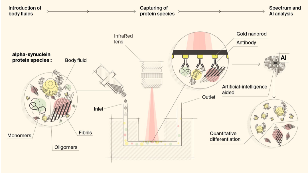
Hatice Altug, Hilal Lashue, and EPFL colleagues have developed ImmunoSEIRA, an AI-enhanced, biosensing tool for the detection of misfolded proteins linked to Parkinson’s and Alzheimer’s disease. The researchers also claim that neural networks can quantify disease stage and progression.

The technology holds promise for early detection, monitoring, and assessing treatment options.

Protein misfolding has been identified as a key event in disease progression. It is thought that healthy proteins misfold first into oligomers , and then into fibrils in later stages.

According to Lashuel, “unlike current biochemical approaches which rely on measuring the levels of these molecules, our approach is focused on detecting their abnormal structures. This technology also allows us to differentiate the levels of oligomers and fibrils.”

The sensor uses gold nanorod arrays with antibodies for specific protein detection, enabling real-time capture and structural analysis of target biomarkers from very small samples. Neural networks identify the presence of specific misfolded protein forms. Lashuel believes that “since the disease process is tightly associated with changes in protein structure, we believe that structural biomarkers, especially when integrated with other biochemical and neurodegeneration biomarkers, could pave the way for more precise diagnosis and monitoring of disease progression.”


Join ApplySci at MIT on September 18, 2023, for a scientist-led day of AI, Deep Tech Health , and Neurotech

Categories
BCI Brain

Brain-spine interface allows paraplegic man to walk

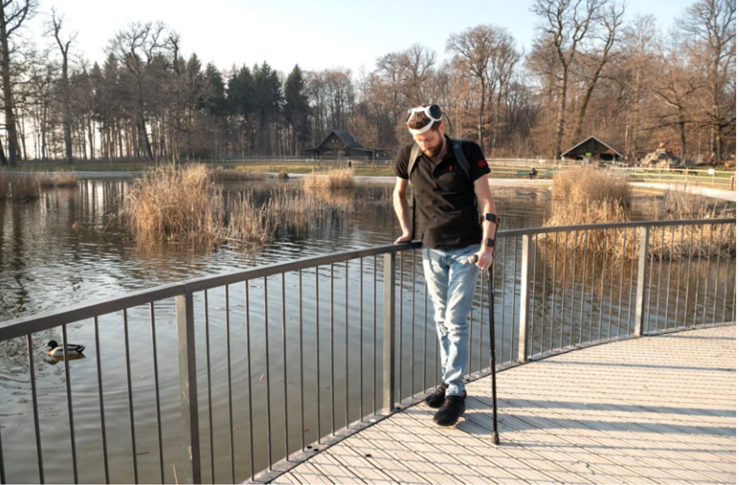
EPFL professor Grégoire Courtine has created a “digital bridge” which has allowed a man whose spinal cord damage left him with paraplegia, to walk.

The brain–spine interface builds on previous work, which combined intensive training and a lower spine stimulation implant. Gert-Jan Oskam participated in this trial, but stopped improving after three years. The new system pairs the existing implant with two disc-shaped skull implants, with two 64-electrode grids resting against the membrane covering the brain.

When Oskam thinks about walking, the skull implants detect electrical activity in the cortex, and wirelessly decode and transmit it to a computer in his backpack, activating the spinal pulse generator.

The previous device “pre-programmed stimulation” that generated robotic stepping movements. Now, Oskam has full control over the parameter of stimulation, allowing him to stop, walk, and climb stairs.

After 40 rehabilitation sessions with the brain–spine interface, Oskam regained the ability to voluntarily move his legs and feet, which was not possible with the previous implant. This suggest that the new device prompted further recovery in nerve cells that were not completely severed during his injury. Oskam can also walk short distances without the device, using crutches.

Courtine is now researching the ability of a similar device to restore arm movement.

Categories
Brain Pharmaceuticals Ultrasound

Implanted ultrasound allows powerful chemotherapy drugs to cross the blood brain barrier

Adam Sonaband and Northwestern colleages used a skull-implantable ultrasound device to open the blood-brain barrier and repeatedly permeate critical regions of the human brain, to deliver intravenous chemotherapy to glioblastoma patients.

This is the first study to successfully quantify the effect of ultrasound-based blood-brain barrier opening on the concentrations of chemotherapy in the human brain. It showed a four- to six-fold increase in concentrations of paclitaxel and carboplatin, powerful drugs not currently used in glioblastoma because they did not cross the blood brain barrier. Temozolomide does cross the blood-brain barrier, but is weaker.

This is also the first study to describe how quickly the blood-brain barrier closes after sonication, allowing optimization of the sequence of drug delivery and ultrasound activation to maximize drug penetration.

The ultrasound implantation takes four minutes, and the patient is awake. It is the basis of an ongoing phase 2 clinical trial.

Categories
Aging Brain

Study: Molecular mechanism of accelerated cognitive decline in women with Alzheimer’s

Hermona Soreq, Yonatan Loewenstein, and Hebrew University of Jerusalem colleagues have uncovered a sex-specific molecular mechanism leading to accelerated cognitive decline in women with Alzheimer’s disease.

Current therapeutic protocols are based on structural changes in the brain and aim to delay symptom progression. Women typically experience more severe side effects from these drugs.

This research shows that severe depletion of mitochondrial RNA fragments inherited from the mother, in the affected brain nuclei, correlates with the rapid deterioration of cognitive abilities in women with living Alzheimer’s.

Soreq explained: “Our research presents a significant contribution to the existing body of Alzheimer’s research by uncovering new insights into the factors driving accelerated cognitive decline in women, underscoring crucial distinctions not only in disease progression but also in treatment response.  Moreover, these findings have implications for treating these symptoms by RNA-based therapies, which emerged in recent years, and now present a viable option.”

Categories
Brain Pharmaceuticals

Donanemab slowed memory decline by 35%, disease progression by 39%, in Alzheimer’s trial

Patients who received Eli Lily’s monthly donanemab infusion in an 18-month study demonstrated a 35% slower decline in memory, thinking, and ability to perform activities of daily living, and were 39% less likely to progress to the next stage of the disease. Brain plaque was also reduced significantly.

The risk of the drug is brain swelling and bleeding. Three trial participants died from these complications.

Lilly CSO/CMO Daniel Skovronsky said donanemab demonstrated the highest level of efficacy of any Alzheimer’s treatment in a clinical trial.

The company plans to apply for FDA approval in the coming months.

Categories
Brain

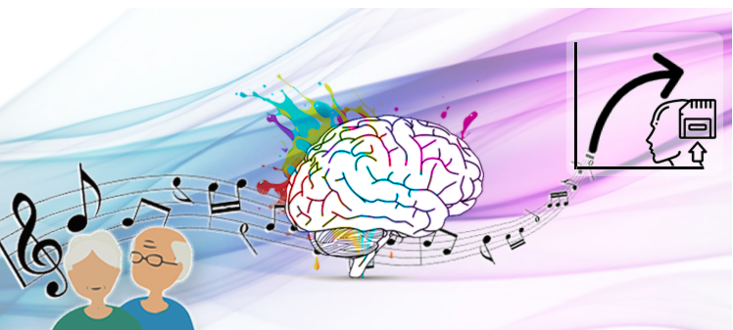
Music improves working memory in study of seniors

A study led by Damian Marie and UNIGE, HES-SO Geneva, and EPFL colleagues showed the effect of music on working memory decline.

132 healthy retirees from 62 to 78 years of age, who had not taken any lessons for at least six months, were assigned to two groups — piano practice, and active listening.

According to author Clara James: ”After six months, we found common effects for both interventions. Neuroimaging revealed an increase in grey matter in four brain regions involved in high-level cognitive functioning in all participants, including cerebellum areas involved in working memory. Their performance increased by 6% and this result was directly correlated to the plasticity of the cerebellum.” Quality of sleep, the number of lessons, and daily training time also had a positive impact on brain performance.

In the pianists, the volume of grey matter remained stable in the right primary auditory cortex. It decreased in the active listening group. A global brain pattern of atrophy was present in all participants, showing that while music can prevent aging in specific regions, the researchers could not yet conclude that music generally rejuvenates the brain.

Categories
Brain Sensors

Closed-loop sensor/stimulation system to detect, reduce neurological events

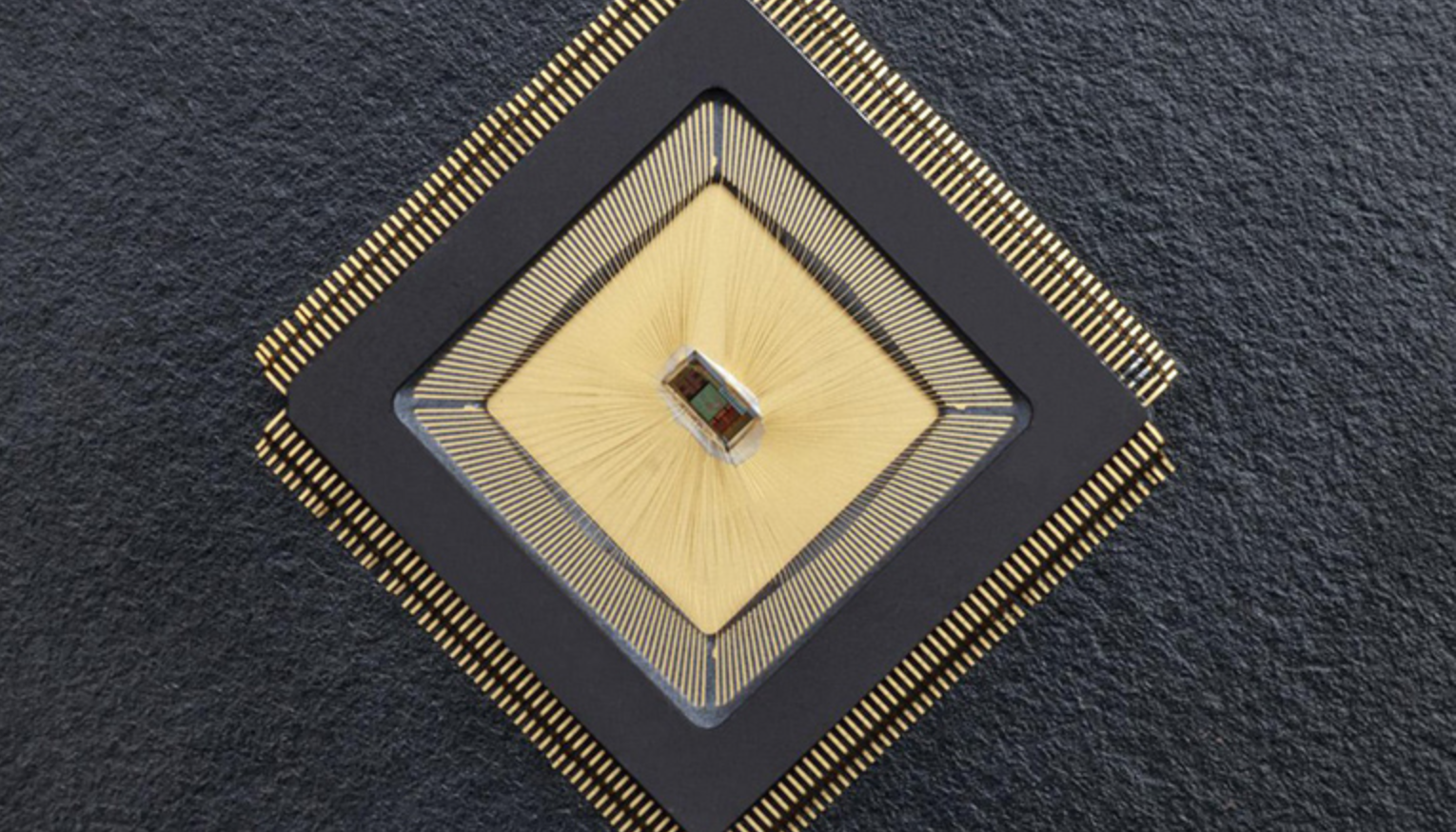
For the first time, there is an autonomous sensing and stimulating unit sitting in a specific brain region and ensuring that neuronal activity in that region is controlled. The closed-loop system is called NeuralTree and was developed by Mahsa Shoaran and EPFL colleagues, to overcome the lack of control caused by epileptic activity or tremor, and later depression, anxiety, OCD, and other disorders. In the future, the technology will be miniaturized, and could rely on internally harvested energy, such as blood flow or oxygen. It could then be possible to implant multiple units to aid the brain, when and where needed.

Categories
AI Brain

AI reconstructs viewed images

Yu TakagiShinji Nishimoto and Osaka University colleagues have published a  study which demonstrates that AI can read brain scans and re-create largely realistic versions of images a person has seen. Future applications could include enabling communication of people with paralysis, recording dreams, and understanding animal perception, among others.

Additional training was used on the existing text-to-image generative AI Stable Diffusion system, linking text descriptions about thousands of photos to brain patterns elicited when those photos were observed. Stable Diffusion was able to get more out of less training for each participant by incorporating photo captions into the algorithm.

The algorithm uses information gathered from regions of the brain involved in image perception, such as the occipital and temporal lobes. The system interpreted information from fMRI scans, detecting changes in blood flow to active brain regions. When people look at a photo, the temporal lobes register information about image contents (people, objects, or scenery), and the occipital lobe predominantly registers information about layout and perspective, such as the scale and position. This is recorded by the fMRI as it captures peaks in brain activity, and these patterns can then be reconverted into an imitation image using AI.

Categories
Brain

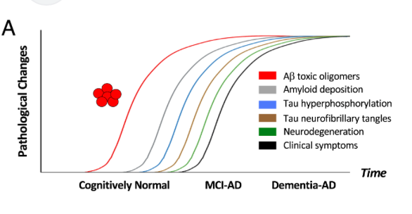
Amyloid beta oligomer blood test could predict Alzheimer’s disease several years in advance

University of Washington’s Valerie Daggett, Dylan Shea, and colleagues, have developed a lab test that can measure levels of amyloid beta oligomers in blood samples. Known as SOBA, the test detected, in a study of 310 subjects, oligomers in the blood of Alzheimer’s patients, but not in most of the control group, which had no cognitive impairment at the time of sample.

SOBA detected oligomers in the blood of 11 individuals from the control group. 10 were diagnosed, years later, with mild cognitive impairment or brain pathology consistent with Alzheimer’s disease. The exam record of the 11th member of the control group was not available.

SOBA (soluble oligomer binding assay) exploits a unique property of the toxic oligomers. When misfolded amyloid beta proteins begin to clump into oligomers, they form a structure known as an alpha sheet. At the heart of SOBA is a synthetic alpha sheet that can bind to oligomers in samples of cerebrospinal fluid or blood. The test then uses standard methods to confirm that the oligomers attached to the test surface are made up of amyloid beta proteins.

UW spinout company AltPep is working to develop SOBA into a diagnostic test for oligomers. In the study, the team also showed that SOBA easily could be modified to detect toxic oligomers of proteins associated with Parkinson’s disease and Lewy body dementia.

Categories
Brain Parkinson's

qMRI for early detection of Parkinson’s disease

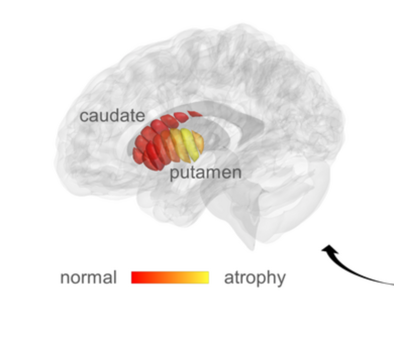
Aviv Mezer and Hebrew University colleagues used quantitative MRI to identify cellular changes in Parkinson’s disease. Their method enabled them to look at microstructures in the striatum, which is known to deteriorate during disease progression. Using a novel algorithm developed by Elior Drori, biological changes in the striatum were revealed, and associated with early stage Parkinson’s, and movement dysfunction.

qMRI achieves its sensitivity by taking several MRI images using different excitation energies. The researchers used this to reveal changes in the tissue structure within distinct regions of the striatum. Previously, the structural sensitivity of these measurements could only be seen post mortem. 

Mezer’s goal is to facilitate early diagnosis of the disease, and provide markers for monitoring the efficacy of future therapies. He also seeks to identify subgroups within the population suffering from Parkinson’s disease – who may respond differently to some drugs than others, leading to personalized treatment. He will now use the technique to investigate microstructural changes in other regions of the brain. The team is in the early stages of developing a qMRI into a tool that can be used in a clinical setting.


Join ApplySci at MIT on September 30 for Deep Tech Health + Neurotech Boston

Categories
Brain

Non-invasive stimulation improves memory in study

In a recent study, Boston University professor Robert Reinhart used tACS to stimulate brain activity in 150 people aged 65-88, resulting in memory improvements for one month.

Stimulating the dorsolateral prefrontal cortex improved long-term memory, while stimulating the inferior parietal lobe, with low-frequency electrical currents, boosted working memory.

Participants were asked to recall 20 words that were read aloud. They underwent tACS for the entire 20 minute duration of the task.After four consecutive days, participants who received high-frequency stimulation of the dorsolateral prefrontal cortex had an improved ability to remember words from the beginning of the lists, which depends on long-term memory. Low-frequency stimulation of the inferior parietal lobe enhanced participants’ recall of items later in the lists, which involves working memory. Performance improved over the four days — and the gains persisted a month later. Those who had the lowest levels of general cognitive function before the study experienced the largest memory improvements.

Changing frequencies and brain regions (applying high-frequency stimulation to the parietal lobe, for instance), or using a ‘sham’ protocol in which the electrical currents were applied only briefly at the beginning and end of the task to mimic the sensation of brain stimulation, did not boost memory.

The team is exploring the use of tACS in Alzheimer’s disease, as the study indicated that brain stimulation might provide the greatest benefits to those who have poor cognitive function.


Join ApplySci at MIT for Deep Tech Health + Neurotech Boston on September 30th, with talks by:

Giovanni Traverso – Bob Langer – Emery Brown – George Church – Mary Lou Jepsen – Tom Oxley – Ellen Roche – Nathan Intrator – Bakul Patel – Ramita Tandon – Shaun Patel – Elizabeth Andukowich – Cris De Luca – Robert Garber – Jonathan Behr – Ann DeWitt