Categories
3-D Printing Brain

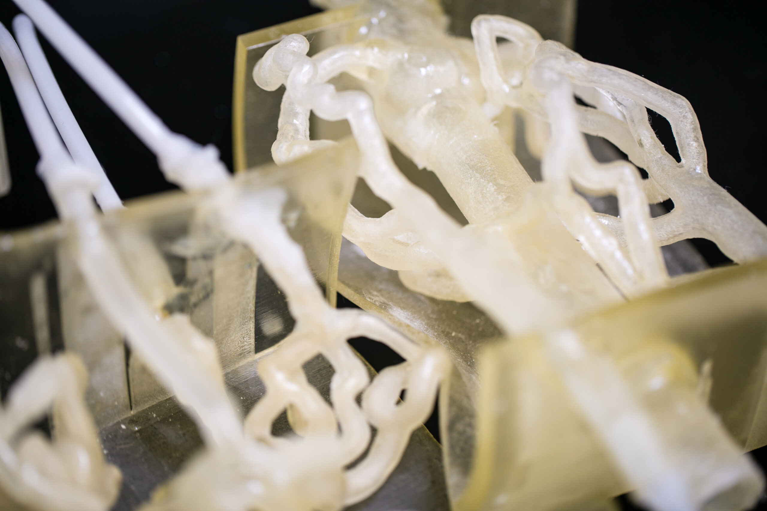
3D printed model for brain aneurysm surgery planning

Stratasys and the Jacobs Institute have used 3D printing for brain surgery planning in an effort to reduce risk. Anatomical models of a patient’s entire brain vessel anatomy were 3D printed before she underwent an aneurysm procedure.

The replica, built of a polymer that mimics human tissue, allowing the surgeons to plan their approach and practice the operation, was based on CT scans.

In this case. the accurate model enabled surgeons to fine-tune the procedure.  “While we were doing that mock procedure, we realized that we had to change some of the tools we wanted to use, given her anatomy,” said  Adnan Siddiqui, Jacobs’ Chief Medical Officer.

WEARABLE TECH + DIGITAL HEALTH SAN FRANCISCO – APRIL 5, 2016 @ THE MISSION BAY CONFERENCE CENTER

NEUROTECH SAN FRANCISCO – APRIL 6, 2016 @ THE MISSION BAY CONFERENCE CENTER
Categories
3-D Printing Heart

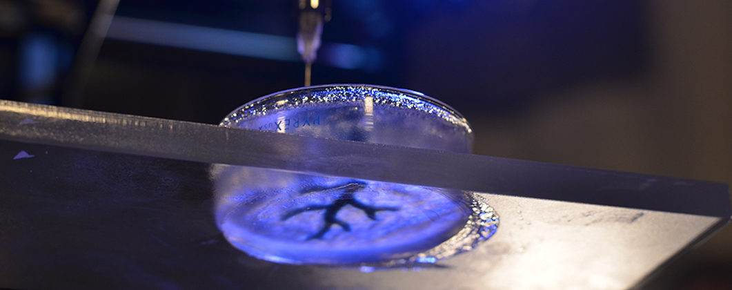
Toward a 3D printed heart

Carnegie Mellon‘s Adam Feinberg is developing 3D printing techniques that could in the future be used to repair the heart.  This work is aimed at alternative solutions for the 4,000 Americans currently waiting to receive a heart transplant.

Feinberg described his progress:  “We’ve been able to take MRI images of coronary arteries and 3-D images of embryonic hearts and 3-D bioprint them with unprecedented resolution and quality out of very soft materials like collagens, alginates and fibrins.”

The next step is to incorporate real heart cells into these 3-D printed tissue structures, providing a scaffold to help form contractile muscle.

Click to view Carnegie Mellon video.

WEARABLE TECH + DIGITAL HEALTH SAN FRANCISCO – APRIL 5, 2016 @ THE MISSION BAY CONFERENCE CENTER

NEUROTECH SAN FRANCISCO – APRIL 6, 2016 @ THE MISSION BAY CONFERENCE CENTER
Categories
3-D Printing Heart

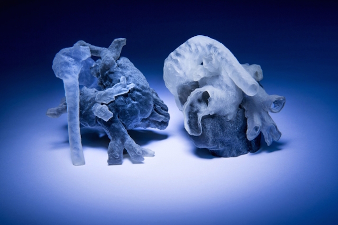
Faster, personalized, 3D printed heart models for surgery planning

MIT and Boston Children’s Hospital researchers are converting heart MRI scans into 3D printed physical models,  for surgical planning,  in 3-4 hours.  Previously, the process took 10 hours. The project, which limits human input to increase accuracy, is led by Professor Polina Golland.  Physicist Medhi Moghari enhanced the precision of the MRI, decreasing the dependence on generic models, and enabling the the team to create the algorithm and print the model in the shorter time frame.

The algorithm examines patches of unsegmented cross sections and looks for similar features in the nearest segmented cross sections. Golland believes that its performance might be improved if it also examined patches that ran obliquely across several cross sections, which will be the next phase of research.

Categories
3-D Printing

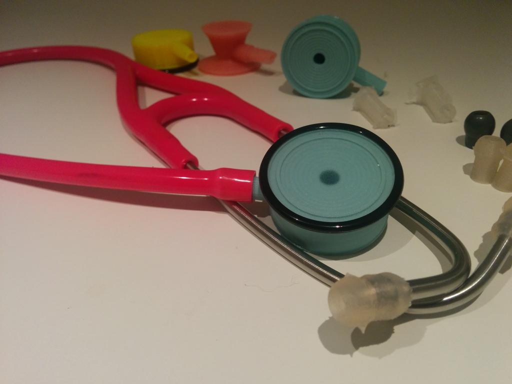
Cheap, accurate, 3D printed stethoscope

Dr. Tarek Loubani has created a 3D printed stethoscope that can be made for $2.50 – $5.00.  Stethoscopes usually cost $150 and are often not available in poor regions.

Through his Glia Project, Dr. Loubani aims to provide cheap, accurate medical supplies, including stethoscopes, electrocardiograms, and pulse oximeters,  to places in need.

“This is simple, cheap and it’s enough for us here,” said Dr. Ayman Sahbani, head of the emergency department at Gaza’s Shifa Hospital, who tested the Glia stethoscope. “Now we can make a stethoscope available for each doctor.”

Categories
3-D Printing Cancer

Cancer patient receives 3D printed rib cage

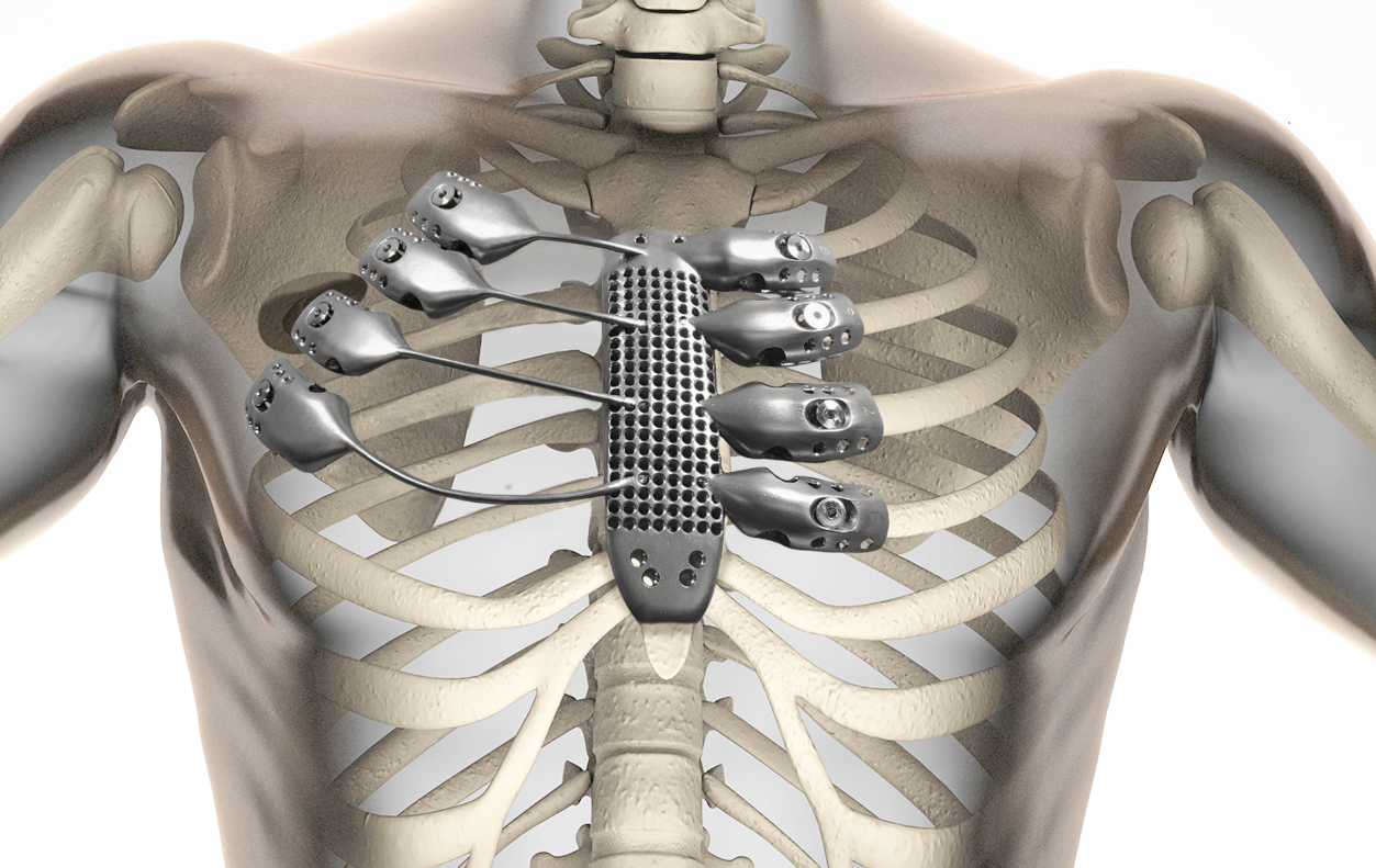
For the first time, a chest wall sarcoma patient has received a  fully customized 3d printed sternum and rib cage portion, created using high resolution CT data.

This part of the chest is difficult to recreate with traditional prosthetics.  Thoracic surgeons typically use flat and plate implants for the chest, which can loosen over time and increase complications.  Rapidly prototyped 3D printed ribs may become the future standard.

View CSIRO video here.

Categories
3-D Printing Respiratory

3D printed airway splints restore breathing

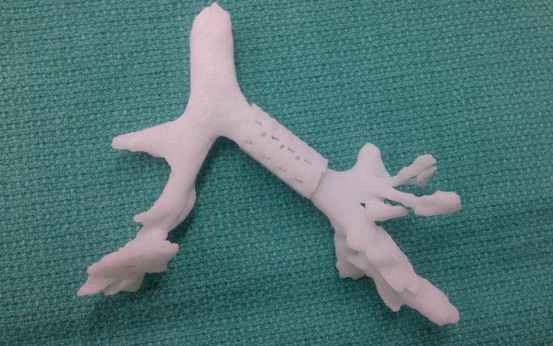
At the University of Michigan, three children under 2 with tracheobronchomalacia had 3D printed devices implanted to open their airways and restore their breathing.

Professors Glenn Green and Scott Hollister were able to create and implant customized tracheal splints for each patient. The device was created directly from CT scans of their tracheas, integrating an image-based computer model with laser-based 3D printing to produce the splint.

The splint was sewn around the patient’s airways to expand the trachea and bronchus and give it a skeleton to aid proper growth. It is designed to be reabsorbed by the body over time. The growth of the airways were followed with CT and MRI scans, and it was shown to allow airway growth for all three patients.

The findings suggest that early treatment of tracheobronchomalacia may prevent complications of conventional treatment such as a tracheostomy, prolonged hospitalization, mechanical ventilation, cardiac and respiratory arrest, food malabsorption and discomfort. None of the devices implanted in this study have caused complications.

The bioresorable splints enabled the patients to come off of ventilators and ended their need for paralytics, narcotics and sedation.  Researchers noted improvements in multiple organ systems.  The patients were also relieved of immunodeficiency-causing proteins that prevented them from absorbing food so that they no longer needed intravenous therapy.

WEARABLE TECH + DIGITAL HEALTH NYC 2015 – JUNE 30 @ NEW YORK ACADEMY OF SCIENCES.  REGISTER HERE.

Categories
3-D Printing

3-D printed organs interlaced with blood vessels

MIT Technology Review

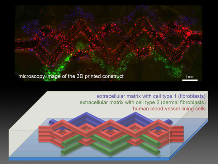
Harvard professor Jennifer Lewis has created a patch of tissue containing skin cells and biological structural material interwoven with blood-vessel-like structures using a 3-D printer and “disappearing” ink.

Lewis’s team created hollow, tube-like structures within a mesh of printed cells using an “ink” that liquefies as it cools. The tissue is built by the 3-D printer in layers. A gelatin-based ink acts as extracellular matrix—the structural mix of proteins and other biological molecules that surrounds cells in the body. Two other inks contained the gelatin material and either mouse or human skin cells. All these inks are viscous enough to maintain their structure after being laid down by the printer.

A third ink with counterintuitive behavior helped them create the hollow tubes. This ink has a Jell-O-like consistency at room temperature, but when cooled it liquefies. The team printed tracks of this ink amongst the others. After chilling the patch of printed tissue, the researchers applied a light vacuum to remove the special ink, leaving behind empty channels within the structure. Then cells that normally line blood vessels in the body can be infused into the channels.

The smallest channels printed were about 75 micrometers in diameter, which is much larger than the tiny capillaries that exchange nutrients and waste throughout the body. The hope is that the 3-D printing method will set the overall architecture of blood vessels within artificial tissue and then smaller blood vessels will develop along with the rest of the tissue.