
 http://spectrum.ieee.org/biomedical/imaging/a-wiring-diagram-of-the-brain
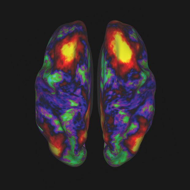
Images of 68 brains from the Human Connectome Project recently became available. The process was powered by highly advanced brain scanning hardware and state of the art image processing and analysis software.
To provide multiple perspectives on each brain, researchers employed several methods:
1. MRI scans provided basic structural images of the brain, providing very high resolution images of the convoluted folds of the cerebral cortex.
2. fMRI scans detected blood flow throughout the brain and showed brain activity for subjects both at rest and engaged in seven different tasks (including language, working memory, and gambling exercises).
3. Diffusion MRI tracked the movement of water molecules within brain fibers. Because water diffuses more rapidly along the length of the fibers that connect neurons than across them, this technique allows researchers to directly trace connections between sections of the brain.
Each imaging modality has its limitations, so combining them gives neuroscientists the best view of how the brain works. The data was purged of noise and artifacts, and then organized into a database.
 http://news.yale.edu/2013/05/07/research-news-real-time-brain-feedback-can-help-people-overcome-anxiety
 http://www.latimes.com/health/boostershots/la-sci-pain-measure-fmri-20130409,0,5200467.story
 http://www.technologyreview.com/news/512141/the-brain-activity-map/