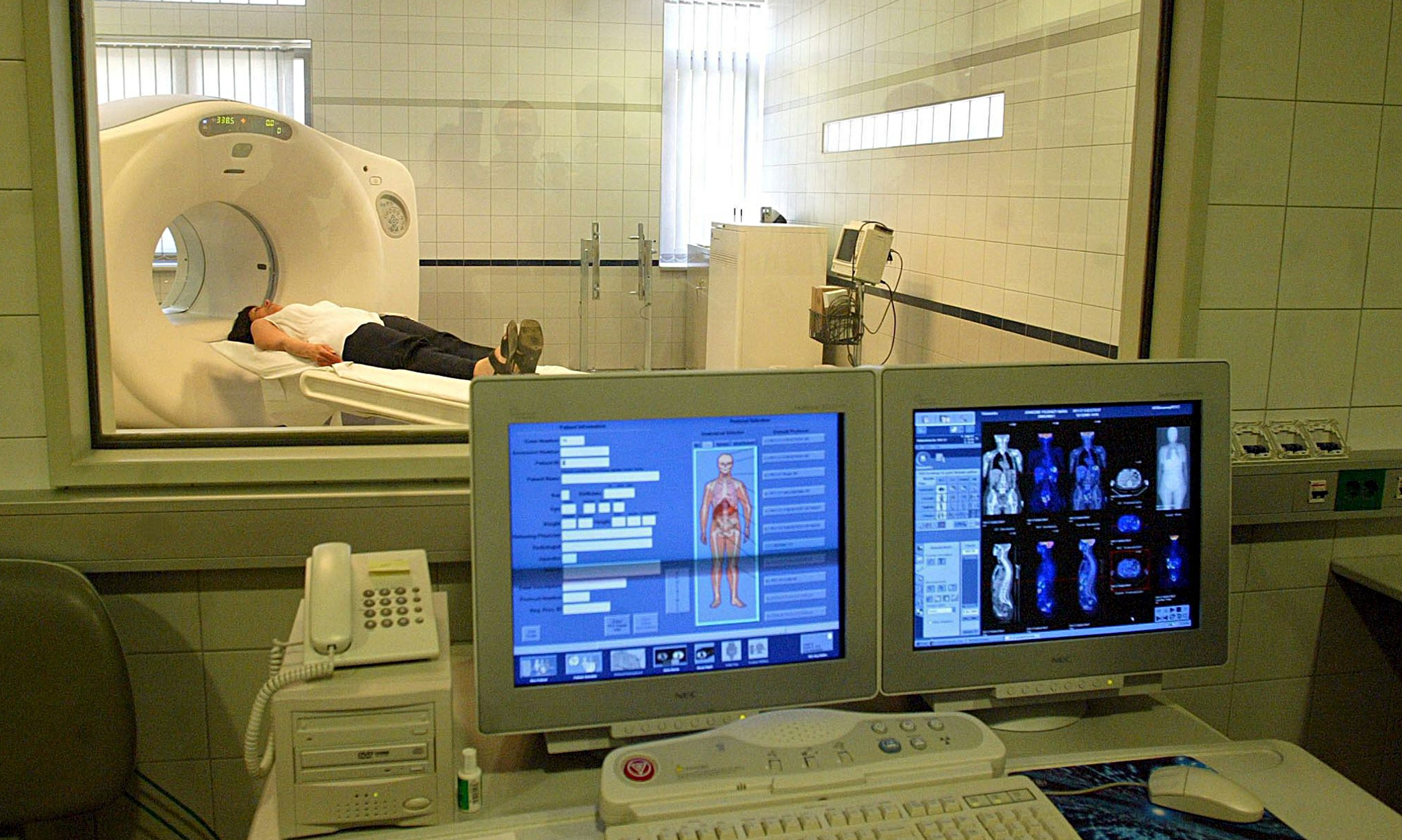
Category: fMRI
-
Brain architecture linked to consciousness, abstract thought
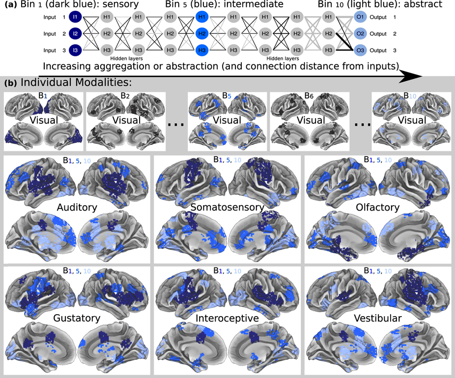
UMass professor Hava Siegelmann used fMRI data from tens of thousands of patients to understand how thought arises from brain structure. This resulted in a geometry-based method meant to advance the identification and treatment of brain disease. It can also be used to improve deep learning systems, and her lab is now creating a “massively recurrent deep learning…
-
Brain scans for customized treatment
MIT‘s John Gabrieli is investigating the use of neuroimaging to predict future behavior to customize brain health treatments. Professor Gabrieli believes that neuromarkers, determined by fMRI, can be used to develop personalized interventions to improve education, health, addiction, criminal behavior and to analyze responses to drug or behavioral treatments. According to Gabrieli, “Presently, we often wait for failure,…
-

Brain imaging technique identifies autism
Virginia Tech Carilion Research Institute professor P. Read Montague has developed a brain imaging technique that may be able to identify autism in children. Current diagnosis is a long an unquantifiable process based on clinical judgment. The study demonstrates that a perspective tracking response can be used to determine whether someone has autism spectrum disorder. It investigates…
-
Transparent, implantable sensor improves brain monitoring
On October 23rd, ApplySci described Justin Williams‘s graphene based, transparent sensor brain implant. The Nature paper is now available online. This will redefine neural implants as it will enable better fMRI monitoring of the activity around the implant, while getting detailed activity from the area. Together with noninvasive EEG, this can help fine tune very detailed EEG…
-
Brain network map may improve non-invasive stimulation
Brain stimulation treatments can alter neural circuits electrically instead of chemically. However, understanding what brain regions should be targeted, by condition, remains a challenge, particularly in non-invasive rTMS. A Beth Israel Deaconess study suggests that brain networks – the interconnected pathways that link brain circuits to one another– can help guide site selection for brain…
-
PET and fMRI predict brain injury recovery
University of Liège professor Steven Laureys‘ recent study shows that PET scans fMRI were more reliable predictors of brain injury recovery than standardized bedside assessments by doctors. Of 126 patients in the trial, 41 were in a persistent vegetative state, 81 were in a minimally conscious state and 4 had locked-in syndrome. PET correctly predicted the extent of recovery…
-
Computational modeling and fMRI show the roles of brain regions in behavioral control
https://www.cell.com/neuron/abstract/S0896-6273(13)01125-2 John O’Doherty of the Caltech Brain Imaging Center has pinpointed areas of the brain—the inferior lateral prefrontal cortex and frontopolar cortex—that seem to serve as the “arbitrator” between model-based and model-free decision-making systems, weighing the reliability of the predictions each makes and then allocating control accordingly. Professor O’Doherty believes that this can lead to better…
-
fMRI shows emotional reactions in vegetative patients
http://www.plosone.org/article/info:doi/10.1371/journal.pone.0074711 Using fMRI, Tel Aviv University and Sourasky Medical Center’s Haggai Sharon, Yotam Pasternak, Talma Hendler and colleagues have shown that the brains of patients in a vegetative state emotionally react to photographs of people they know, as though they recognize them. “We showed that patients in a vegetative state can react differently to different stimuli in the…
-
Imaging technology identifies signs of chronic brain injury in living football players
http://www.technologyreview.com/news/522126/identifying-signs-of-chronic-brain-injury-in-living-football-players/ Until now, chronic traumatic encephalopathy, caused by repetive head injury, could only be identified after a victim died. CTE is linked to depression, dementia, and memory loss. A new imaging method, developed by Pittsburgh Steelers physician Julian Bailes and UCLA researchers, can for the first time spot signs of the condition in the living brain. It…
-
Brain imaging method improves resolution in PAG studies
http://www.pnas.org/content/early/2013/09/25/1306095110.abstract The “midbrain periaqueductal gray region,” or PAG, is extraordinarily difficult to investigate in humans because of its size and intricate structure. Northeastern University researcher Ajay Satpute is uses state-of-the art imaging to capture this complex neural activity. His technique increases the spatial resoluion of fMRI. As fMRI lacks temporal resolution, there is much room for improvement. Satpute’s goal…
-
Neurofeedback enhances signal-to-noise ratio in thought
http://research.vtc.vt.edu/news/2013/sep/13/covert-operations-your-brain-digitally-remastered/. Virgina Tech Carillon Professor Stephen Laconte developed technology to transfer non-invasive brain activity measurements into control signals that drive physical devices and computer displays in real time. The study suggests that the signal-to-noise ratio of the brain activity underlying our thoughts can be remastered. Researchers used whole-brain, classifier-based real-time fMRI to understand the neural underpinnings…
-
fMRI and machine learning identify emotions
http://www.plosone.org/article/info%3Adoi%2F10.1371%2Fjournal.pone.0066032 Carnegie Mellon researchers have developed “a new method with the potential to identify emotions without relying on people’s ability to self-report” using a combination of fMRI and machine learning. They recruited 10 actors from the university’s drama school to act out emotions including anger, happiness, pride and shame, while inside an fMRI scanner, multiple times in…