 http://stm.sciencemag.org/content/5/201/201ra119
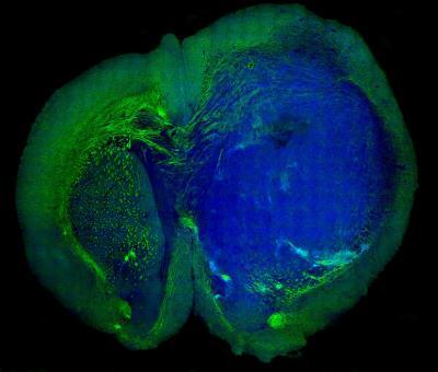
Harvard’s Xiaoliang Sunney Xie and Minbiao Ji used SRS microscopy (Stimulated Raman Scattering) to “see” the tiniest areas of tumor cells in brain tissue, and to distinguish tumor from healthy tissue in the brains of living mice. They then showed that the same was possible in tissue removed from a patient with glioblastoma multiforme, one of the most deadly brain tumors.
Professor Xie described that “Biopsy has been the gold standard for detecting and removing these types of tumors. But this technique, we believe, is better because it’s live. Surgeons can now skip all the steps of taking a biopsy, freezing, and staining the tissue. This technique allows them to do it all in vivo.”
SRS works by shining non-invasive lasers into tissue and detecting the weak signal that emerges. By analyzing the signal’s spectrum, researchers can build images of the cellular makeup of the tissue. By amplifying those signals, they transform a technique that once took hours or days into one that works in real time, and could offer a critical insight to surgeons in the operating room. Since brain tissue and tumors contain different chemical makeups, researchers can create images that precisely show where the tumor “margin” — the boundary area where tumor cells infiltrate among normal cells — is located, helping to guide surgeons in the operating room.
Leave a Reply