Alan Jasanoff and MIT colleagues are using MRI to monitor calcium activity at a much deeper level in the brain than previously possible, to show how neurons communicate with each other. The research team believes that this enables neural activity to be linked with specific behaviors.
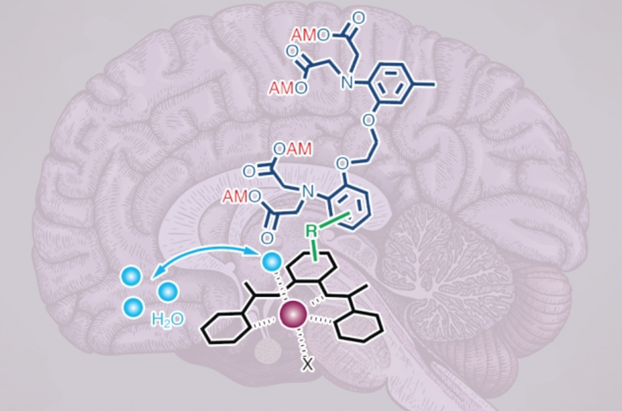
To create their intracellular calcium sensors, the researchers used manganese as a contrast agent, bound to an organic compound that can penetrate cell membranes, containint a calcium-binding chelator.
Once inside the cell, if calcium levels are low, the calcium chelator binds weakly to the manganese atom, shielding the manganese from MRI detection. When calcium flows into the cell, the chelator binds to the calcium and releases the manganese, which makes the contrast agent appear brighter in an MRI.
The technique could also be used to image calcium as it performs in facilitating the activation of immune cells, or in diagnostic brain or heart imaging.
Join ApplySci at the 12th Wearable Tech + Digital Health + Neurotech Boston conference on November 14, 2019 at Harvard Medical School and the 13th Wearable Tech + Neurotech + Digital Health Silicon Valley conference on February 11-12, 2020 at Stanford University