Brown University‘s Diane Hoffman-Kim, Yu-Ting Dingle and Molly Boutin have developed a cheap method for developing a 3D mini brain for biomedical research.
The central nervous system tissue sphere can produce electrical signals and form synapses. Applications include drug testing, neural tissue transplant testing, and stem cell experiments.
The mini-brains are not the first or most sophisticated working cell cultures of a central nervous system, but they require fewer steps to make and use readily available materials. Dingle compares the technology to retail 3-D printers, which have proliferated, bringing once-rare technology to a mass market. “We could allow all kinds of labs to do this research,” she said.
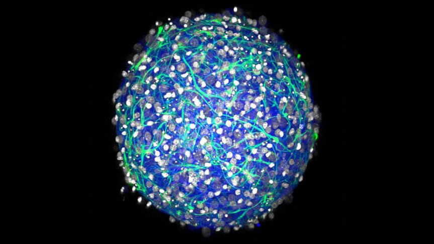
The method yields mini-brains with several properties:
- Diverse cell types: The cultures contain both inhibitory and excitatory neurons and several varieties of essential neural support cells called glia.
- Electrically active: the neurons fire and spike and form synaptic connections, producing complex networks.
- 3-D: Cells connect and communicate within a realistic geometry, rather than merely across a flat plane as in a 2-D culture.
- Natural density: Experiments showed that the mini-brains have a density of a few hundred thousand cells per cubic millimeter, which is similar to a natural rodent brain.
- Physical structure: Cells in the mini-brain produce their own extracellular matrix, producing a tissue with the same mechanical properties (squishiness) as natural tissue. The cultures also don’t rely on foreign materials such as scaffolds of collagen.
- Longevity: In testing, cultured tissues live for at least a month.
The spheres of brain tissue begin to form within a day after the cultures are seeded, and form complex 3-D neural networks in 2-3 weeks.
NEUROTECH SAN FRANCISCO CONFERENCE – APRIL 6, 2016 – EARLY REGISTRATION ENDS OCTOBER 17TH.