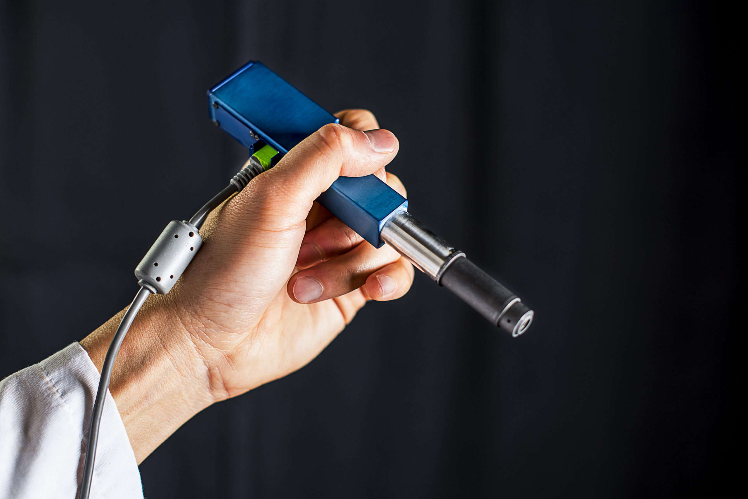
University of Washington, Memorial Sloan Kettering Cancer Center, Stanford University and Barrow Neurological Institute researchers are developing a handheld, miniature microscope to allow surgeons to “see” at a cellular level in the operating room. This can enable more precise brain tumor removal, as surgeons try not to leave cancerous material behind, while protecting healthy brain matter.
According to lead author Jonathan Liu, “Surgeons don’t have a very good way of knowing when they’re done cutting out a tumor. They’re using their sense of sight, their sense of touch, pre-operative images of the brain — and oftentimes it’s pretty subjective. Being able to zoom and see at the cellular level during the surgery would really help them to accurately differentiate between tumor and normal tissues and improve patient outcomes,”
The microscope delivers high-quality images at faster speeds than existing devices. In addition to tumor surgery, researchers will begin testing it as a cancer-screening tool in dental and dermatological clinics, in an effort to avoid invasive biopsies and waiting time at labs.
Wearable Tech + Digital Health San Francisco – April 5, 2016 @ the Mission Bay Conference Center
NeuroTech San Francisco – April 6, 2016 @ the Mission Bay Conference Center
Wearable Tech + Digital Health NYC – June 7, 2016 @ the New York Academy of Sciences
NeuroTech NYC – June 8, 2016 @ the New York Academy of Sciences