Categories
AI Brain Cancer

AI decodes brain tumor DNA during surgery

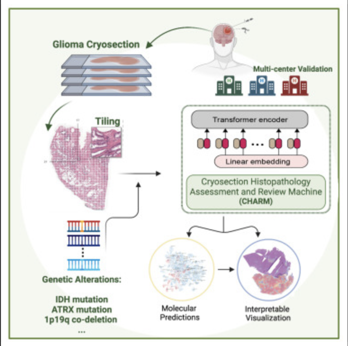
Kun-Hsing Yu and HMS colleagues used AI to rapidly determine a brain tumor’s molecular identity during surgery, propeling the development of precision oncology. The tool is CHARM (Cryosection Histopathology Assessment and Review Machine.) Currently, genetic sequencing takes days to weeks.  

Accurate molecular diagnosis during surgery can help a neurosurgeon decide how much brain tissue to remove. Removing too much when the tumor is less aggressive can affect a patient’s neurologic and cognitive function. Removing too little when the tumor is highly aggressive may leave behind malignant tissue that can grow and spread quickly. 

The technology will also allow the surgeon to determine if the patient can benefit from immediate treatment with drug-coated wafers placed directly into the brain at the time of the operation.

The standard intraoperative diagnostic approach used now involves taking brain tissue, freezing it, and examining it under a microscope. A major drawback is that freezing the tissue tends to alter the appearance of cells under a microscope and can interfere with the accuracy of clinical evaluation. Furthermore, the human eye, even when using potent microscopes, cannot reliably detect subtle genomic variations on a slide.

The new AI approach overcomes these challenges and could be particularly valuable in areas with limited access to technology to perform rapid cancer genetic sequencing.

Knowledge of a tumor’s molecular type provides insight about its aggressiveness, behavior, and likely response to various treatments, which can inform post-operative decisions.

The new tool enables during-surgery diagnoses aligned with the WHO classification system for diagnosing and grading the severity of gliomas, which calls for such diagnoses to be made based on a tumor’s genomic profile.

CHARM was developed using 2,334 brain tumor samples from 1,524 people with glioma from three different patient populations. When tested on a never-before-seen set of brain samples, the tool distinguished tumors with specific molecular mutations at 93 percent accuracy and successfully classified three major types of gliomas with distinct molecular features that carry different prognoses and respond differently to treatments.

It successfully captured visual characteristics of the tissue surrounding the malignant cells. It was capable of spotting telltale areas with greater cellular density and more cell death within samples, both of which signal more aggressive glioma types.

CHARM was also able to pinpoint clinically important molecular alterations in a subset of low-grade gliomas, a subtype of glioma that is less aggressive and therefore less likely to invade surrounding tissue. Each of these changes also signals different propensity for growth, spread, and treatment response.

It further connected the appearance of the cells — the shape of their nuclei, the presence of edema around the cells — with the molecular profile of the tumor. This means that the algorithm can pinpoint how a cell’s appearance relates to the molecular type of a tumor.

Accorging to Yu, this ability to assess the broader context around the image renders the model more accurate and closer to how a human pathologist would visually assess a tumor sample.

The researchers said that while the model was trained and tested on glioma samples, it could be successfully retrained to identify other brain cancer subtypes. 

Scientists have already designed AI models to profile other types of cancer — colon, lung, breast — but gliomas have remained particularly challenging due to their molecular complexity and huge variation in tumor cells’ shape and appearance.

The CHARM tool would have to be retrained periodically to reflect new disease classifications as they emerge from new knowledge. “Just like human clinicians who must engage in ongoing education and training, AI tools must keep up with the latest knowledge to remain at peak performance,” according to Yu.


Join ApplySci at MIT on September 18, 2023 for the 14th AI + Deep Tech Health + Neurotech conference.
Categories
Cancer Ultrasound

Wearable ultrasound to detect early breast cancer

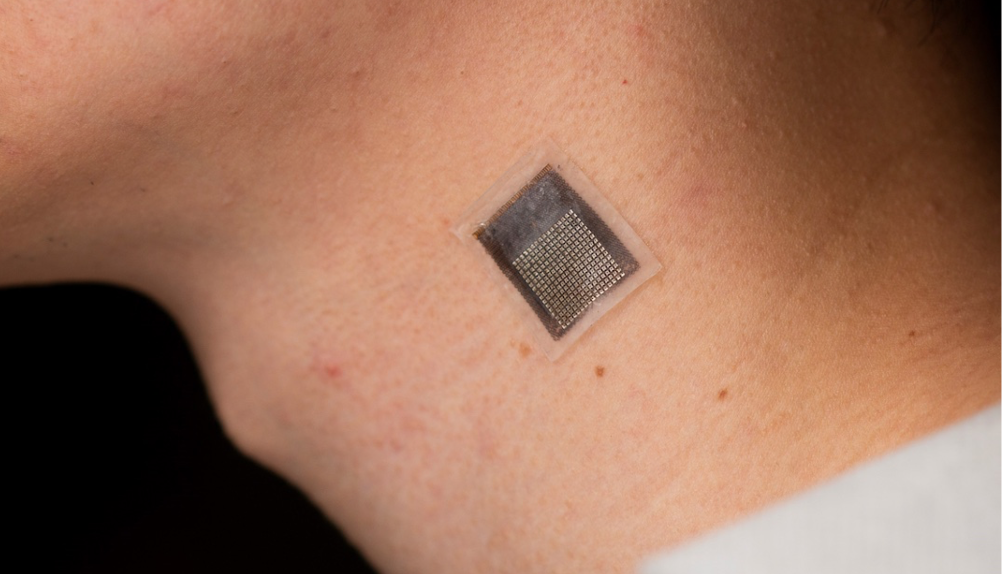
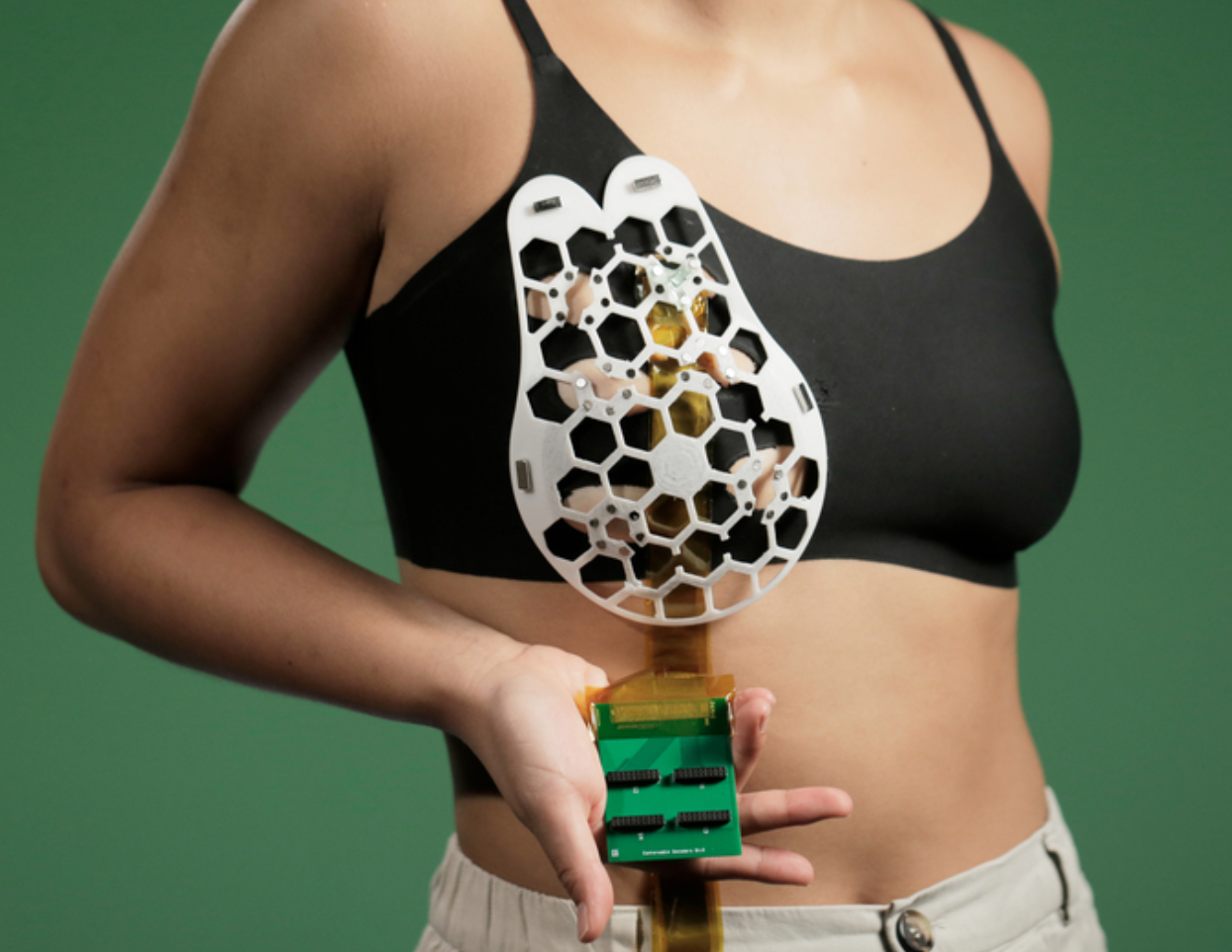
MIT’s Canan Dagdeviren has developed a flexible ultrasound patch that can be attached to a bra, obtaining ultrasound images with resolution comparable to medical imaging centers, and used repeatedly.

Interval cancers, which develop between regularly scheduled mammograms, account for 20 – 30 percent of all breast cancers, and tend to be more aggressive. The goal is to frequently screen those most likely to develop interval cancers.

According to Dagdeviren: “We changed the form factor of the ultrasound technology so that it can be used in your home. It’s portable and easy to use, and provides real-time, user-friendly monitoring of breast tissue.”

Piezoelectric material allowed the scanner to be minimized, in a flexible, 3D-printed patch, with honeycomb shaped openings. Using magnets, it is attached to a bra with openings that allow the it to contact the skin. The scanner fits inside a small tracker, with six different positions, allowing the entire breast to be imaged. IT also rotates, to take images from different angles.

The device was able to detect .3cm diameter cysts in a 71-year-old woman, with a resolution comparable to that of traditional ultrasound. Tissue was imaged at a depth up to 8 centimeters.

To see the images, the scanner must connect to an ultrasound machine, like those used in imaging centers. The team is now working on a miniaturized imaging system.


Join ApplySci at MIT on September 18, 2023 for AI + Deep Tech Health + Neurotech Boston

Categories
AI Cancer

AI-based device increases ademona detection during colonoscopy

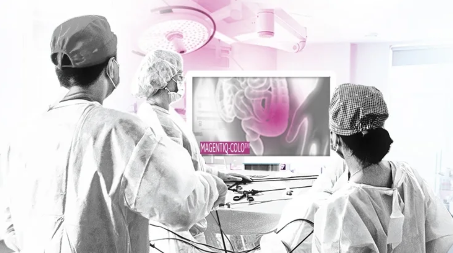
MAGENTIQ-COLO is an AI based FDA cleared colonoscopy device which offers a significant increase in Adenoma Detection Rate.

Current high rates of missed and undetected adenomas during colonoscopy means that even regularly screened patients are still at risk of developing colon cancer. A missed polyp can lead to interval cancer, which accounts for approximately 8% to 10% of all CRC in the U.S., translated to over 13,500 cancer cases that could be prevented every year with better detection.

A 2022 study of 950 patients at 10 hospitals showed that MAGENTIQ-COLO increasing ADR by 26% relatively, translating to a 21% decrease in CRC occurrence and a 35% decrease in patient mortality.


Join ApplySci at MIT on September 18, 2023 for AI + Deep Tech Health + Neurotech Boston

Categories
AI Cancer

AI predicts pancreatic cancer

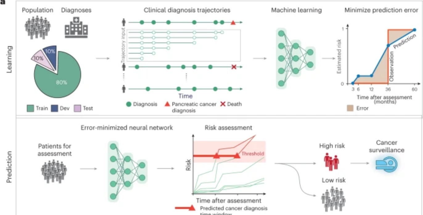
Harvard professor Chris Sander used clinical data from 6 million patients in Denmark’s national health system and 3 million in the U.S. VA system to train an AI model to predict the occurrence of pancreatic cancer within 3, 6, 12, and 36 months. This could allow wider screening for the aggressive disease, which is often discovered at a late stage, including in people with no known genetic risk.

The researchers believe that data from imaging, genetic, and wearable devices could further improve this tool, and that unrelated disease histories, including diabetes and substance abuse have already improved its accuracy.


Join ApplySci at MIT for the 14th AI+ Deep Tech + Neurotech conference on the future of healthcare on September 18, 2023

Categories
Cancer Heart Sensors

Wearable sensor evaluates human tissue stiffness

Sheng Xu and colleagues have developed a wearable, stretchable device that non-invasively evaluates the stiffness of human tissue, at an improved penetration depth, and for a longer period than, existing methods.

An ultrasonic array facilitates serial, non-invasive, three-dimensional imaging of tissues, four centimeters below the surface of human skin, at a spatial resolution of 0.5 millimeters.

The sensor can be used to detect cancer progression, which cases cells to stiffen; diagnose and treat sports injuries, by monitoring muscles, ligaments and tendons; and monitor the efficacy of treatments for liver, cardiovascular disease, and cancer, which cause tissue to stiffen.

Categories
AI Cancer

AI catches breast cancer earlier, more often than traditional screening alone

The mammography screening paradigm has not changed since the 1960s.

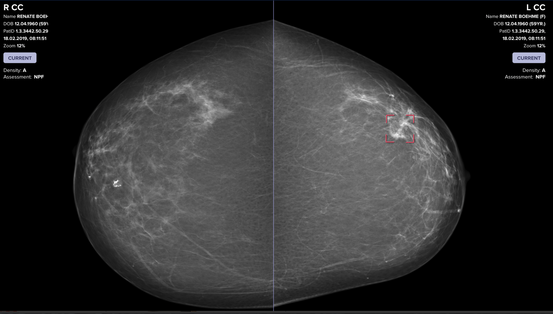
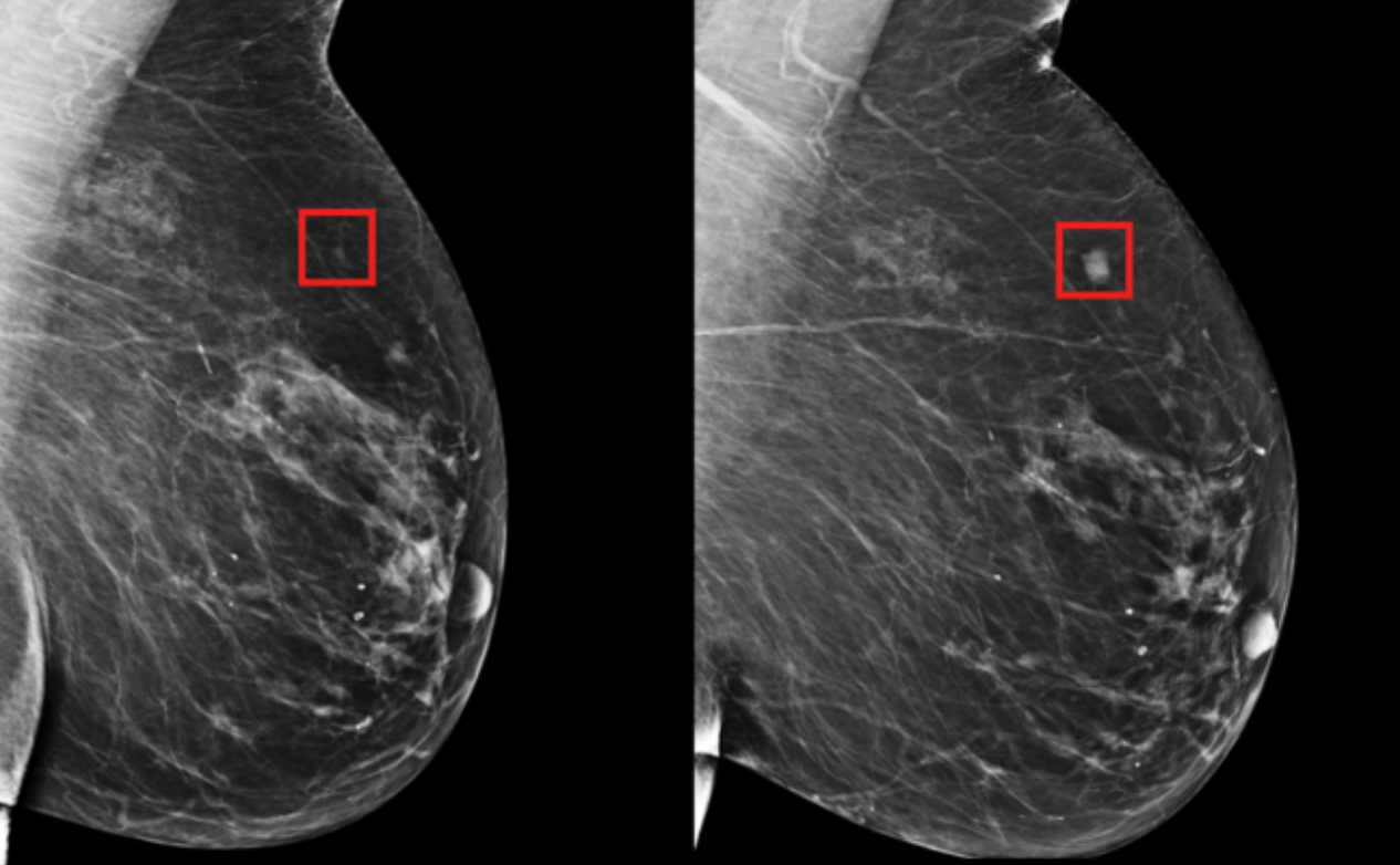
Breast screening AI company Vara, with Essen University and Memorial Sloan Kettering hospitals, published a study showing that radiologists assisted by AI are better able to screen for breast cancer. The hope is that AI systems could detect cancers that doctors miss, provide better care in remote areas, and allow radiologists more time to see more patients.

Two approaches were tested. In the first, AI analyzed mammograms. In the second, AI distinguished between normal and concerning scans. It refers the latter to a radiologist, who reviews them before seeing the AI’s assessment. Then the AI issues a warning if it detected cancer when the doctor did not. In the study, the AI examined old scans and compared its assessments with those of the radiologist who reviewed them.

Data from 367,000 mammograms—including radiologists’ notes, original assessments, and information on whether the patient ultimately had cancer— was analyzed to learn how to place these scans into one of three categories: “confident normal,” “not confident” (in which no prediction is given), and “confident cancer.” The conclusions from both approaches were then compared with the decisions real radiologists originally made on 82,851 mammograms sourced from screening centers that didn’t contribute scans used to train the AI.

The second approach—doctor and AI working together—was 2.6% better at detecting breast cancer than a doctor working alone, and raised fewer false alarms. It automatically set aside scans it classified as “confident normal,” which was 63% of all mammograms.


Clairity, an AI-driven precision breast cancer screening company, using Harvard professor Connie Lehman and colleagues’ decades of research in radiomics and applied AI, has developed a rigorous scientific approach to improve the accuracy of risk assessment. Prof Lehman discussed her approach at the 2019 ApplySci conference at Harvard Medical School.


Join ApplySci at MIT on September 30, 2022 for Deep Tech Health + Neurotech Boston featring Giovanni Traverso, MITBakul Patel, GoogleGeorge Church, Harvard Kerri Dugan, DARPAEllen Roche, MITNathan Intrator, NeurosteerEmery Brown, HarvardMary Lou Jepsen, OpenwaterShaun Patel, REACT NeuroElizabeth Andukowich, NIMHRamita Tandon, WalgreensCris de Luca, Sanofi VenturesTom Oxley, Synchron

Categories
Cancer Sensors

MSK developed sensor detects molecular signature of cancer; compared to human scent

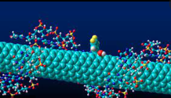
Mijin Kim and Daniel Heller of the Nanomedicine Lab at Memorial Sloan Kettering Cancer Center have developed an array of carbon nanotube sensors that can “sniff” cancer using AI.

The human nose can detect a trillion different scents, through hundreds of olfactory receptors. The pattern which odor molecules bind to which receptors creates a kind of molecular signature that the brain uses to recognize a scent.

Like the nose, the cancer detection technology uses an array of multiple sensors to detect a molecular signature of the disease, interpreted by machine learning.

Each nanotube sensor can detect many different molecules in a blood sample. By combining the many responses of the sensors, the technology creates a unique fluorescent pattern. The pattern can be recognized by an algorithm trained to identify the difference between a cancer fingerprint and a normal one.

In experiments conducted on ovarian cancer patient blood, the nanosensor detected ovarian cancer more accurately than current biomarker tests. The researchers believe that the technique could be adapted to detect multiple types of cancer using the same set of sensors without first identifying biomarkers.


Join ApplySci at MIT on September 30th for Deep Tech Health + Neurotech Boston

Categories
AI Cancer

AI for (much) earlier breast cancer detection – Constance Lehman

Connie Lehman — Professor, Radiology, Harvard Medical School; Chief of Breast Imaging, radiology, Massachusetts General Hospital; Co-director of AVON Breast Center, Radiology, Massachusetts General Hospital; and Director, Breast Imaging Research Center, Radiology, Massachusetts General Hospital spoke at ApplySci’s recent conference at Harvard Medical School.

Click to listen to her talk on using AI to discover breast cancer 5 years earlier than currently possible.


Join ApplySci at the 13th Wearable Tech + Digital Health + Neurotech Silicon Valley conference on February 11-12, 2020 at Quadrus Sand Hill Road.  Speakers include:  Zhenan Bao, Stanford – Vinod Khosla, Khosla Ventures – Mark Chevillet, Facebook – Shahin Farshchi, Lux Capital – Carla Pugh, Stanford – Nathan Intrator, Tel Aviv University | Neurosteer – Wei Gao, Caltech – Sergiu Pasca, Stanford – Walter Greenleaf, Stanford – Sheng Xu, UC San Diego – Dror Ben-Zeev, University of Washington – Mikael Eliasson, Roche  – Unity Stoakes, StartUp Health

Categories
Cancer

PET scan tracer detects both cancer and lung disease

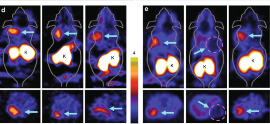
Stanford’s Sanjiv Gambhir has developed an imaging molecule that can identify pancreatic, cervical and lung cancer early– and, surprisigly, hard-to-detect idiopathic pulmonary fibrosis.  The tracer clings to integrin alpha-v beta-6. In a PET scan, the tracer glows due to radioactivity emissions, which allows doctors to see exactly where the tracer is docked in the body.

A small clinical trial included patients healthy and cancer or IPF patients. The results indicated the tracer is accurate and has disease detection potential.


Join ApplySci at the 12th Wearable Tech + Digital Health + Neurotech Boston conference on November 14, 2019 at Harvard Medical School featuring talks by Brad Ringeisen, DARPA – Joe Wang, UCSD – Carlos Pena, FDA  – George Church, Harvard – Diane Chan, MIT – Giovanni Traverso, Harvard | Brigham & Womens – Anupam Goel, UnitedHealthcare  – Nathan Intrator, Tel Aviv University | Neurosteer – Arto Nurmikko, Brown – Constance Lehman, Harvard | MGH – Mikael Eliasson, Roche – Nicola Neretti, Brown – R. Jacob  Vogelstein, Camden Partners

Join ApplySci at the 13th Wearable Tech + Neurotech + Digital Health Silicon Valley conference on February 11-12, 2020 on Sand Hill Road featuring talks by Zhenan Bao, Stanford – Rudy Tanzi, Harvard – Shahin Farshchi – Lux Capital – Sheng Xu, UCSD – Carla Pugh, Stanford – Nathan Intrator, Tel Aviv University | Neurosteer – Wei Gao, Caltech – Mikael Eliasson, Roche – Dror Ben-Zeev, University of Washington – Sergiu Pasca, Stanford

Categories
Cancer

Infrared light detects tumors under the skin

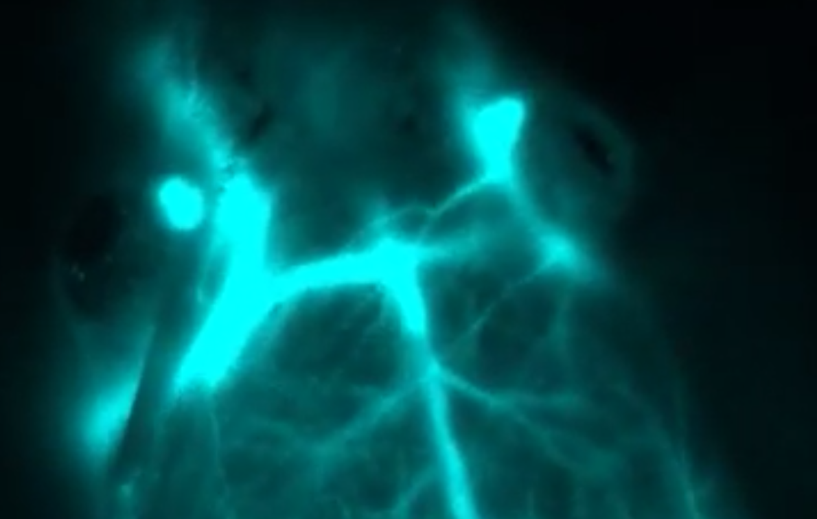
Stanford’s Hongjie Dai has developed a deep-tissue imaging technique that clearly illuminates tumors beneath the skin.  It relies on nanoparticles containing erbium,  which glows in the infrared.  The promising technology has only been tested on mice, so far.

In a study, the technique was used to predict cancer patient response to immunotherapy, and to measure drug response and tumor size after treatment.

Researcher Zhuoran Ma said:  “Our approach allows for seeing into an intact mouse brain while conventional approaches see only the scalp”

Researcher Yeteng Zhong said:, “The combined imaging depth, molecular specificity and multiplicity, and spatial and temporal resolution are unattainable by previous techniques.”

This could provide a noninvasive way to identify candidates for drugs with out a biopsy.

Click to view Stanford video


Join ApplySci at the 12th Wearable Tech + Digital Health + Neurotech Boston conference on November 14, 2019 at Harvard Medical School featuring talks by Brad Ringeisen, DARPA – Joe Wang, UCSD – Carlos Pena, FDA  – George Church, Harvard – Diane Chan, MIT – Giovanni Traverso, Harvard | Brigham & Womens – Anupam Goel, UnitedHealthcare  – Nathan Intrator, Tel Aviv University | Neurosteer – Arto Nurmikko, Brown – Constance Lehman, Harvard | MGH – Mikael Eliasson, Roche – Nicola Neretti, Brown

Join ApplySci at the 13th Wearable Tech + Neurotech + Digital Health Silicon Valley conference on February 11-12, 2020 on Sand Hill Road featuring talks by Zhenan Bao, Stanford – Rudy Tanzi, Harvard – Shahin Farshchi – Lux Capital – Sheng Xu, UCSD – Carla Pugh, Stanford – Nathan Intrator, Tel Aviv University | Neurosteer – Wei Gao, Caltech

Categories
AI Cancer

AIgorithm detects cancer potential of pancreatic cysts

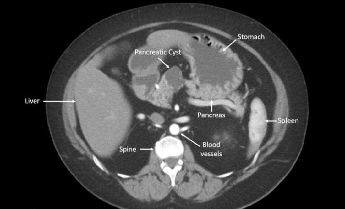
CompCyst is a proof-of-concept study, led by Anne Marie Lennon at Johns Hopkins, which uses AI to more accurately determine which pancreatic cysts will become cancerous. The test evaluates molecular and clinical markers in cyst fluids, and could significantly improve detection rates vs. current clinical and imaging tests.

In the study, the researchers evaluated molecular profiles, including DNA mutations and chromosome changes, of 862 pancreatic cysts. An algorithm developed by David Masica classified patients into the three groups: those with no potential to turn cancerous, for which patients would not require periodic monitoring; mucin-producing cysts that have a small risk of progressing to cancer, for which patients can receive periodic monitoring for progression to possible cancer; and cysts for which surgery is recommended because there is a high likelihood of progression to cancer.

Based on histopathological analysis of surgically resected cysts, the researchers found that surgery was not needed in 45% of patients. This unnecessary surgery was performed because the clinicians could not determine the cysts were dangerous. If CompCyst had been used, the researchers estimated that 60% to 74% of the patients (depending on the cyst type) could have been spared unnecessary intervention.


Join ApplySci at the 12th Wearable Tech + Digital Health + Neurotech Boston conference on November 14, 2019 at Harvard Medical School featuring talks by Brad Ringeisen, DARPA – Joe Wang, UCSD – Carlos Pena, FDA  – George Church, Harvard – Diane Chan, MIT – Giovanni Traverso, Harvard | Brigham & Womens – Anupam Goel, UnitedHealthcare  – Nathan Intrator, Tel Aviv University | Neurosteer – Arto Nurmikko, Brown – Constance Lehman, Harvard | MGH – Mikael Eliasson, Roche – David Rhew, Samsung

Join ApplySci at the 13th Wearable Tech + Neurotech + Digital Health Silicon Valley conference on February 11-12, 2020 at Stanford University featuring talks by Zhenan Bao, Stanford – Rudy Tanzi, Harvard – David Rhew, Samsung – Carla Pugh, Stanford – Nathan Intrator, Tel Aviv University | Neurosteer

Categories
AI Cancer

Deep learning mammography model detects breast cancer up to five years in advance

MIT CSAIL professor Regina Barzilay and Harvard/MGH professor Constance Lehman  have developed a deep learning model that can predict breast cancer, from a mammogram, up to five years in the future. The model learned subtle breast tissue patterns that lead to malignant tumors from mammograms and known outcomes of 90,000 MGH patients.

The goal is to individualize screening and prevention programs.

Barzilay said that “rather than taking a one-size-fits-all approach, we can personalize screening around a woman’s risk of developing cancer.  For example, a doctor might recommend that one group of women get a mammogram every other year, while another higher-risk group might get supplemental MRI screening.”

The algortithm accurately placed 31 percent of all cancer patients in its highest-risk category, compared to 18 percent for traditional models.

Lehman hopes to change screening strategies from age-based to risk based. “This is because before we did not have accurate risk assessment tools that worked for individual women.”

Current risk assement,  based on age, family history of breast and ovarian cancer, hormonal and reproductive factors, and breast density, are weakly correlated with breast cancer. This makes many organizations believe that risk-based screening is not possible.

Rather than manually identifying the patterns in a mammogram that drive future cancer, the algorithm deduced patterns directly from the data, detecting abnormalities too subtle for the human eye to see.

Lehman said that “since the 1960s radiologists have noticed that women have unique and widely variable patterns of breast tissue visible on the mammogram. These patterns can represent the influence of genetics, hormones, pregnancy, lactation, diet, weight loss, and weight gain. We can now leverage this detailed information to be more precise in our risk assessment at the individual level.”

The MIT/MGH model  is equally accurate for white and black women, as opposed to prior models. Black women have been shown to be 42 percent more likely to die from breast cancer due to a wide range of factors that may include differences in detection and access to health care.

Barzilay believes the system could, in the future,  determine, based on mammograms, if patients are at a greater risk for cardiovascular disease or other cancers.


Professor Constance Lehman will discuss this technology at ApplySci’s 12th Wearable Tech + Digital Health + Neurotech Boston conference on November 14, 2019 at Harvard Medical School