Categories
Brain fMRI Neural Computation

Computational modeling and fMRI show the roles of brain regions in behavioral control

https://www.cell.com/neuron/abstract/S0896-6273(13)01125-2

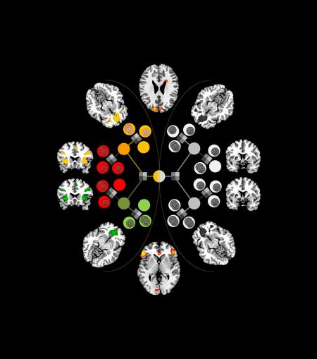
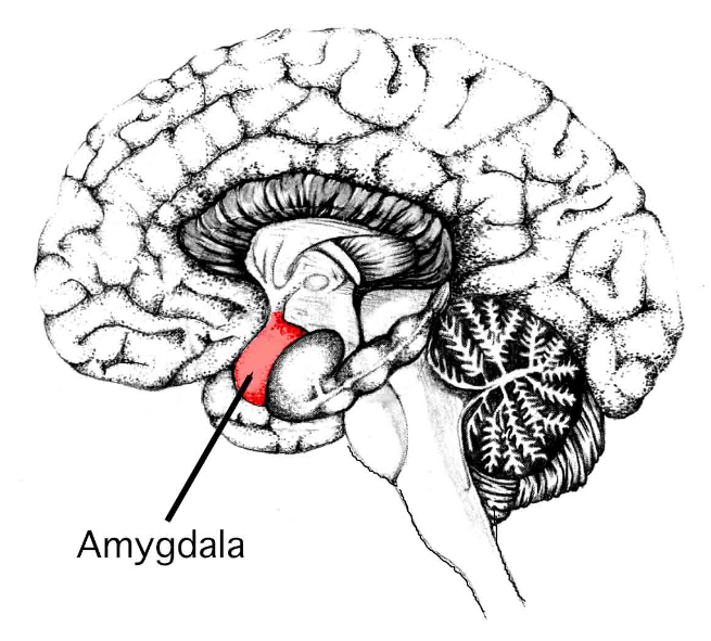
John O’Doherty of the Caltech Brain Imaging Center has pinpointed areas of the brain—the inferior lateral prefrontal cortex and frontopolar cortex—that seem to serve as the “arbitrator” between model-based and model-free decision-making systems, weighing the reliability of the predictions each makes and then allocating control accordingly.  Professor O’Doherty believes that this can lead to better treatments for brain disorders, such as drug addiction, and psychiatric disorders, such as obsessive-compulsive disorder. These disorders, which involve repetitive behaviors, may be driven in part by malfunctions in the degree to which behavior is controlled by the habitual system versus the goal-directed system.

Categories
Assistive Technologies Brain Heart Stroke

Non-invasive, nanoparticle method for identifying atherosclerosis plaques

http://pubs.acs.org/doi/abs/10.1021/nl404816m

Case Western‘s Michael Bruckman and colleagues have developed a multifunctional nanoparticle that pinpoints blood vessel plaques caused by atherosclerosis using MRI.  The goal is to create a non-invasive method of identifying heart attack and stroke causing plaques vulnerable to rupture, in time for treatment.

Currently doctors can only identify narrowing blood vessels caused by plaque accumulation via incision and the insertion of a catheter inside a blood vessel in the arm, groin or neck. The catheter emits a dye that enables X-rays to show the narrowing.

The researchers found that a nanoparticle built from a rod-shaped virus, commonly found on tobacco, locates and illuminates plaque in arteries more effectively, with a fraction of the dye.  The tailored nanoparticles target plaque biomarkers, opening the possibility that particles can be programmed to identify vulnerable plaques from stable.  Untargeted dyes alone cannot accomplish this.

Categories
Prosthetics Robotics Sensors

Trial: Improved sense of touch and control in prosthetic hand

http://stm.sciencemag.org/content/6/222/222ra19

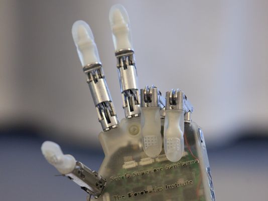
In the ongoing effort to improve the dexterity of prosthetics, a recent trial showed an improved sense of touch and control over a prosthetic hand.  EPFL professor Silvestro Micera and colleagues surgically attached electrodes from a robotic hand to a volunteer’s median and ulnar nerves. Those nerves carry sensations that correspond with the volunteer’s index finger and thumb, and with his pinky finger and the edge of his hand, respectively. The volunteer controlled the prosthetic with small muscle movements detected by sEMG, a non-invasive method that measures electrical signals through the skin.

Over seven days, the volunteer was asked to grasp something with a light grip, a medium grip, and a hard grip, and to evaluate the shape and stiffness of three kinds of objects. During 710 tests, he wore a blindfold and earphones so that he could not use his vision or sound to guide the prosthetic. The researchers also sometimes turned off the sensory feedback to test whether he was using time to modulate his grip.

The subject was able to complete the requested tasks with his prosthetic thumb and index finger 67 percent of the time the first day and 93 percent of the time by the seventh day of the experiment. His pinky finger was harder to control: he was only able to accomplish the requested grip 83 percent of the time. In both the grip strength tests and in detecting the stiffness of objects, the volunteer made mistakes with the medium setting and object, but he never confused the softest and hardest objects. The ability to modulate his grip strength is this study’s main progress over previous work by the same group.

Categories
Cancer Ultrasound

Ultrasound combined with contrasting agent for radiation-free tumor detection

http://www.plosone.org/article/info%3Adoi%2F10.1371%2Fjournal.pone.0086642

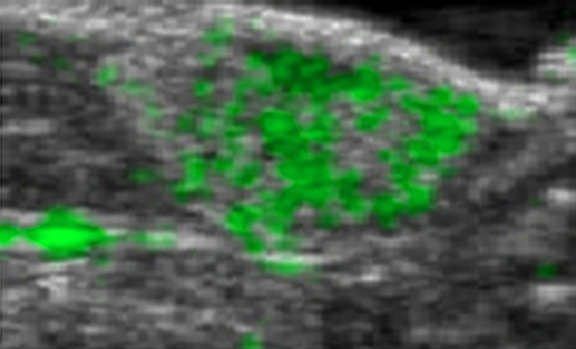
University of North Carolina Professor Nancy Klauber-Demore has improved the resolution and tumor-detecting ability of ultrasound scans.

Combining ultrasound with a contrast agent composed of tiny bubbles that pair with an antibody that many cancer cells produce at higher levels than do normal cells, Klauber-Demore was able to visualize lesions created by angiosarcoma.  By binding to the protein SFRP2, the contrast agent may help distinguish malignant from benign masses found on imaging.  Since SFRP2 is expressed in many cancers – including breast, colon, pancreas, ovarian, and kidney tumors – the technique could potentially be useful for a broad range of cancer types.  As the level of SFRP2 in tumors increases as tumors develop, the team will also investigate whether the technique can be used to track tumor growth.

Categories
Stem Cells

Nobel-worthy stem cell discovery

http://www.nature.com/news/acid-bath-offers-easy-path-to-stem-cells-1.14600

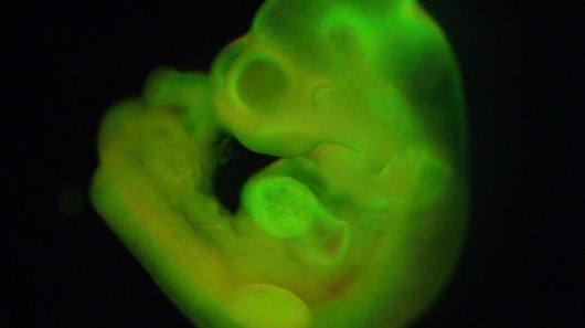
Haruko Obokata and colleagues at the RIKEN Center for Developmental Biology have created embryonic stem cells from a single blood cell by putting white blood cells from a baby mouse in a mild acid solution. Eventually a few stem cells emerge that can turn into any other cell in the body including skin, heart, liver or neurons.

Scientists have long searched for ways to make human embryonic stem cells that did not destroy human embryos. These cells hold great potential for treating Alzheimer’s, Parkinson’s, heart disease and diabetes.

Obokata put the blood cells in a mild acid for about 30 minutes. The pH of the solution was about 5.7. A few days later, the cells stopped acting like blood and started behaving like stem cells.  When the researchers injected the cells into a mouse embryo, the cells acted just like other stem cells: They created all the organs needed for an adult mouse. The team named the cells stimulus-triggered acquisition of pluripotency, or STAP.

This breakthrough could enable scientists to create stem cells from any person, thus controlling genetic similarity, and use them to repair nerve injuries.

Categories
Brain

Fluorescent tags allow researchers to observe molecules becoming memories

https://www.einstein.yu.edu/news/releases/968/watching-molecules-morph-into-memories/

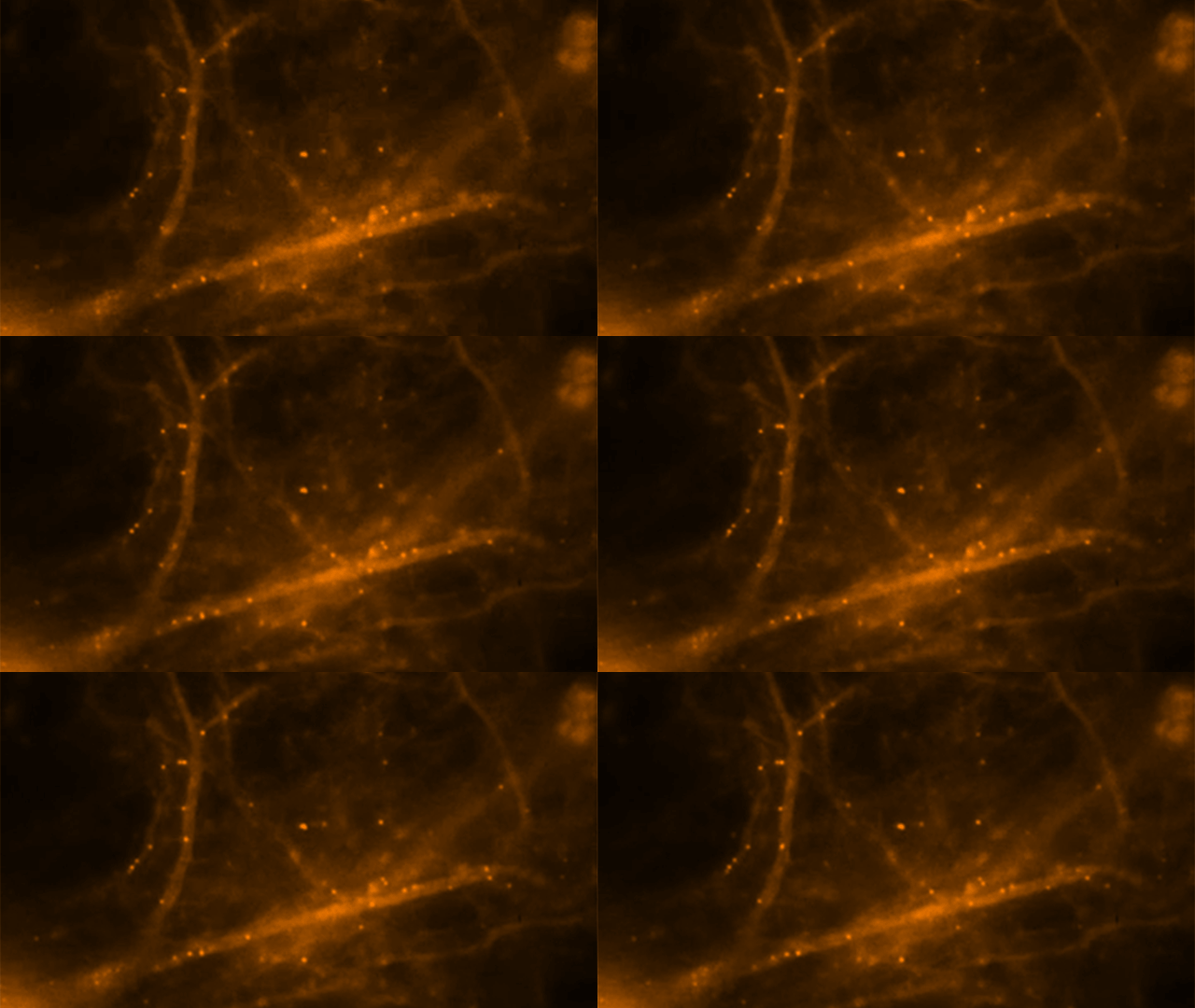
Albert Einstein College of Medicine researchers have used advanced imaging techniques to observe memory making molecules travel in real time in living brain cells.

To look deeply into neurons without harming them, the researchers developed a mouse model in which they fluorescently tagged all molecules of messenger RNA that code for beta-actin protein – an essential structural protein found in large amounts in brain neurons and considered a key player in making memories. mRNA is a family of RNA molecules that copy DNA’s genetic information and translate it into the proteins that make life possible. They stimulated neurons from the mouse’s hippocampus, where memories are made and stored, and then watched fluorescently glowing beta-actin mRNA molecules form in the nuclei of neurons and travel within dendrites, the neuron’s branched projections. They discovered that mRNA in neurons is regulated through a novel process described as “masking” and “unmasking,” which allows beta-actin protein to be synthesized at specific times and places and in specific amounts.

Neurons come together at synapses, where slender dendritic “spines” of neurons grasp each other, much as the fingers of one hand bind those of the other. Evidence indicates that repeated neural stimulation increases the strength of synaptic connections by changing the shape of these interlocking dendrite “fingers.” Beta-actin protein appears to strengthen these synaptic connections by altering the shape of dendritic spines. Memories are thought to be encoded when stable, long-lasting synaptic connections form between neurons in contact with each other.

Hye Yoon Park stimulated individual hippocampal neurons of the mouse and observed newly formed beta-actin mRNA molecules within 10 to 15 minutes, indicating that nerve stimulation had caused rapid transcription of the beta-actin gene. Further observations suggested that these beta-actin mRNA molecules continuously assemble and disassemble into large and small particles, respectively. These mRNA particles were seen traveling to their destinations in dendrites where beta-actin protein would be synthesized.

Adina Buxbaum showed that neurons may be unique among cells in how they control the synthesis of beta-actin protein.  This revealed the mechanism by which brain neurons handle this challenge. She found that as soon as beta-actin mRNA molecules form in the nucleus of hippocampal neurons and travel out to the cytoplasm, the mRNAs are packaged into granules and so become inaccessible for making protein. She then saw that stimulating the neuron caused these granules to fall apart, so that mRNA molecules became unmasked and available for synthesizing beta-actin protein.

Categories
Eyes Monitoring Sensors

Smart holograms diagnose and monitor medical conditions

http://www.cam.ac.uk/research/news/holographic-diagnostics-0

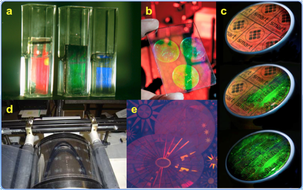
Cambridge researchers are developing responsive, color-changing diagnostic holograms.  Silver nanoparticles are formed into three dimensional holograms of predetermined shapes in a fraction of a second using a single laser pulse.  They will be used for portable medical tests and devices, to monitor diabetes, cardiac function, infections, electrolyte or hormone imbalance easily,inexpensively, and non-invasively.

The ‘smart’ holograms can be used to test blood, breath, urine, saliva or tears for a wide range of compounds, such as glucose, alcohol, hormones, drugs, or bacteria. When one of these compounds is present, the hologram changes color, potentially making the monitoring of various conditions as simple as checking the color of the hologram against a color gradient. Clinical trials of the holographic sensors to monitor glucose levels and urinary tract infections in diabetic patients are currently underway.

Categories
BCI Brain

Neurofeedback alters brain plasticity in PTSD

1. Tel Aviv University paper   2. Schulich School of Medicine paper

Professor Ruth Lanius and colleagues at Western University’s Schulich School of Medicine have published a study showing that neurofeedback can improve subjective wellbeing in PTSD, potentially leading to new treatment options. Schulich researchers claim that this is “the first study to show that key brain networks involved in mediating affect and cognition in PTSD can be volitionally modulated via neurofeedback, with measurable outcomes on subjective well-being. It was achieved by harnessing multiple imaging techniques, including EEG and fMRI. Using fMRI we captured the patients’ resting-state brain activity just before and after a 30-minute neurofeedback training session, which was carried out outside the scanner using EEG. We then searched for any differences in connectivity within well-known brain networks. Interestingly, we discovered significant correlations between EEG and fMRI network activities as well as changes in self-reported calmness. This indicated that neurofeedback was able to directly modulate the brain bases of emotional processing in PTSD.”

A group at the Functional Brain Center at Tel Aviv University is trying to achieve similar results using EEG neurofeedback only.  Yehudit Meir-Hasson, Professor Talma HendlerProfessor Nathan Intrator and colleagues published a recent paper demonstrating the validation of an emotional EEG fingerprint with fMRI in the journal NeuroImage.

Categories
AI BCI Brain Robotics

Collaborative cloud system for human-serving robots

http://www.roboearth.org/what-is-roboearth

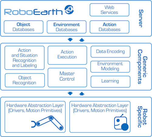
RoboEarth’s goal is to speed the development of human-serving robots. Scientists from five European universities gathered this week for its launch and demonstrated potential applications. This included a robot scanning a room’s physical layout, including the location of the patient’s bed, and the placing a carton of milk on a nearby table.

The system is sometimes called a “Wikipedia” for robots, allowing them, or their programmers,  to share and retrieve information.

Researchers claim that the robots’ technically sophisticated capabilities are comparable to those of high-end robots in car factories. They, however,  look clumsier because robots that interact with humans are not performing repetitive tasks in the controlled, sanitized and predictable surroundings of a factory.

RoboEarth’s system of networked computers enable it to perform intensive computing tasks that smaller computers (or simpler robots) cannot.  It also allows individual robots to communicate between themselves through its “RoboCloud” and robot database. This could facilitate light and cheap robots that use the cloud for computing.

Categories
Eyes Sensors

Miniature 3D sensor maps surgical field; scope small enough for use in neurosurgery

http://www.visionsense.com/hp.html

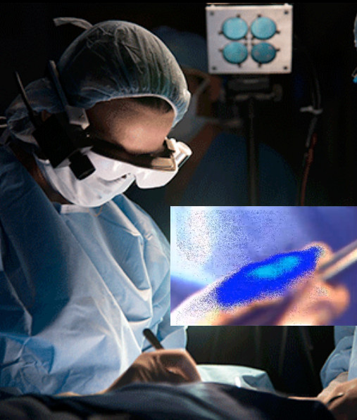
Visionsense’s miniature 3D sensor optically maps the surgical field, based on (and imitating) the eye of a bee. A single sensor is divided into hundreds of thousands of tiny “eyes” looking in different directions, using an array of micron-sized elements.  The elemental information is translated into left and right eye images, resulting in a clear stereoscopic view,  bypassing the diffraction limit that restricts current miniature cameras.

In neurosurgery, scopes must be very small in diameter to pass through narrow ports, such as the nose.  Most 3D scopes rely on two optical channels, each containing a single sensor. Each sensor collects two separate images that are combined to give the appearance of three dimensions as a user looks at the screen – mirroring the sight of the human eye.   Using their single sensor system, Visionsense has developed a high quality image producing scope that is small enough to operate on the brain.

Categories
Diabetes Heart Sensors

Piezoelectric nanoribbons power pacemakers

http://news.cnet.com/8301-11386_3-57617483-76/nanoribbons-let-beating-hearts-power-their-own-pacemakers/

Scientists are studying various steady energy source alternatives for small biomedical sensors, including piezoelectric power.

University of Illinois researchers have attached small, flexible strips (piezoelectric nanoribbons) to internal organs of animals, and harvested energy from their movement  to power pacemakers and other medical devices.  Current practice depends on hard-to-change batteries.

This is a minimally invasive power source, which in the future can be activated by muscle activity other than the heart muscle, with potential implications for glucose pumps and other devices.  The researchers claim that their system is a  “complete, flexible, and integrated system that is capable of harvesting and storing energy from the natural contractile and relaxation motions of the heart, lung, and diaphragm at levels that meet requirements for practical applications.”

Categories
Brain Eyes

Brain processes images viewed for 13 milliseconds

http://web.mit.edu/newsoffice/2014/in-the-blink-of-an-eye-0116.html

MIT professor Mary Potter has published a study showing that the human brain is capable of processing images viewed through the eyes for 13 milliseconds. This is significantly faster than the 100 milliseconds reported in earlier research.

The study offers evidence that “feedforward processing” — the flow of information in only one direction, from retina through visual processing centers in the brain — is enough for the brain to identify concepts without having to do any further feedback processing.  It also suggests that while the images are seen for only 13 milliseconds before the next image appears, part of the brain continues to process those images for longer than that.

The researchers are now investigating how long visual information presented so briefly can be held in the brain. They are also scanning subjects’ brains with a magnetoencephalography (MEG) scanner during the task to see what brain regions are active when a person successfully completes the identification task.