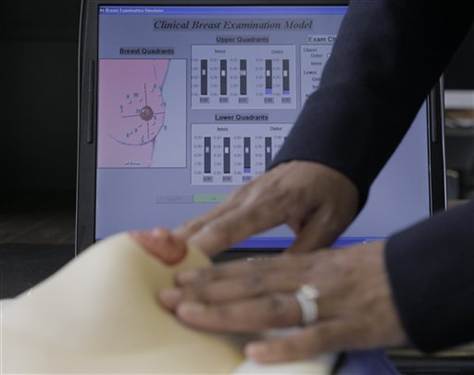
University of Wisconsin‘s Carla Pugh has developed a sensor based breast model to help train physicians to detect tumors. The device indicates when a physician is palpating (pressing) with enough force to detect a lump in the breast. The amount of pressure is displayed as colors on a breast map displayed on a monitor. Blue indicates low pressure and red indicates the highest pressure.
In a study, 53 doctors performed Clinical Breast Exams on 4 sensor enabled breast models, each with a mass of different density located in different areas of the breast. The masses represent potential tumors. Two of the models contained masses near the surface of the breast, and two contained masses at the back of the breast against the chest wall.
Data was collected using video and sensor recordings of the amount of pressure applied by those who found the mass and those who did not. Analysis of the data showed that 15% of the physicians tested were using a technique that did not detect the deep tissue lesions near the chest wall in two of the four breast models.
Wearable Tech + Digital Health NYC 2015 – June 30 @ New York Academy of Sciences. Early registration rate available until 4/24.