Vienna scientists create 3-D human brain tissue from stem cells

http://www.technologyreview.com/news/518716/researchers-grow-3-d-human-brain-tissues/

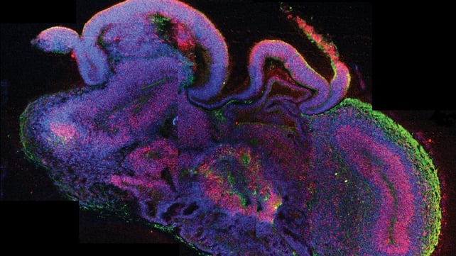
Scientists at the Austrian Academy of Sciences have turned human stem cells into pea-sized mini-brains with a neural structure similar to the brain of a developing embryo.  These “cerebral organoids”, as they are termed formally, are the best living model of a human brain created to date.

The researchers have already used their mini-brains to investigate one neuronal disorder, microcephaly, in which the brain does not grow properly. They hope to apply the technique to more complex conditions such as autism and schizophrenia, for which no good animal models are available.

Posted

in

by

Tags:

Comments

2 responses to “Vienna scientists create 3-D human brain tissue from stem cells”

  1. Dr Manjit Sharma

    This is an amazing advance. How many factors did the stem cells require to “create” or assemble into a whole brain? In what order were these introduced into 3D cultures?Were different genes turned on/off as the brain progressed to regionalisation? Will the research on autism and schizophrenia be carried out on the cortical parts? How will it link to these disorders which manifest in an individual after birth?

  2. Dr Manjit Sharma

    As a postdoc, I had to set up an organ culture in vitro comprising of hypothalamus and hypophysis complex to study feedback regulation of reproductive hormones. I eventually did it and was able to maintain it under sterile conditions in (2D) cultures for 2 days. The tissue looked fine and well maintained, so to say. But when it came to publishing, serious questions were raised about the viability of long term hypothalamic cultures. Even my own doctorate mentor advised me against going ahead because till then only viability of cultured neurons but not brain tissue was acceptable. Viability of cultured hypophysis being glandular was acceptable. I was called upon to carry out several standard histological tests for brain tissue and demonstrate its viability or death before publishing. Eventually it was said that the hypothalamic tissue could be considered to be remarkably well maintained or preserved but not functional.
    Today, seeing this experiment on “cerebral organoid” in 3D cultures has revived memory of my tribulations at the time.
    In view of my earlier experience in 2D cultures I have the following concerns:
    1. Should the brain organoid tissue in 3D cultures be considered as remarkably well preserved or functional?
    2. Have the standard histology tests of neurons/glia, available in the neurology departments of all hospitals, been done to verify its structural integrity?
    3. What tests have been done to demonstrate cerebral organoid’s viability which will allow it to be used in future studies?

Leave a Reply

Your email address will not be published. Required fields are marked *