Judy Wawira Gichoya and Emory colleagues have developed an AI model that detects warning signs for diabetes in x rays collected during routine exams. The signs were also detected in patients who do not meet elevated risk guidelines.
Applying deep learning to images and electronic health record data, the model that successfully flagged elevated diabetes risk in a retrospective analysis, often years before patients were diagnosed.
Current guidelines suggest screening patients for type 2 diabetes if they are between 35 and 70 years old and have a body mass index (BMI) in the overweight to obese range. This strategy misses a significant number of cases, particularly in racial/ethnic minorities for whom BMI is a less effective predictor of diabetes risk.
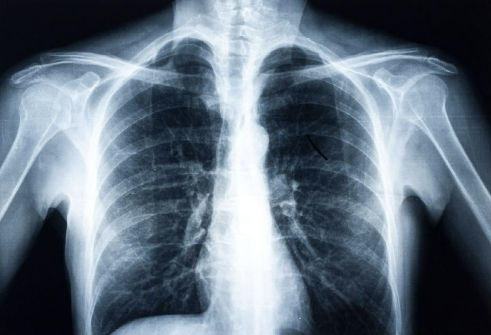
Each year, millions of Americans receive chest x-rays for chest pain, difficulty breathing, injury or before surgeries. While radiologists are not looking for diabetes when they assess these x-rays, the images become part of a patient’s medical record and could be analyzed later for diabetes or other conditions.
The AI model was trained on more than 270,000 x-ray images from 160,000 patients, with deep learning determining the image features that best predicted a later diagnosis of diabetes. Because chest x-rays are not a common way to detect diabetes, the researchers also used explainable AI techniques to determine how and why the model made its determinations. The methods pointed to the location of fatty tissue as important for determining risk, a logic that aligns with recent medical findings that visceral fat in the upper body and abdomen is associated with type 2 diabetes, insulin resistance, hypertension and other conditions.
When the Emory team applied the model to a separate group of nearly 10,000 patients, they found the model predicted risk better than a simple model based on non-image clinical data alone.
In some cases, the chest x-ray warned of high diabetes risk as early as three years before the patient eventually received a diagnosis. The model’s output also provides a numerical risk score that could potentially help clinicians customize the treatment approach for patients
The research team will now explore how to further validate the model and incorporate it into electronic health record systems so it can provide an alert to physicians to pursue traditional diabetes screening of patients flagged as high risk based on x-ray results.
They’ll then turn to investigating how well chest x-rays can help diagnose other conditions, such as vascular disease, congestive heart failure, and chronic obstructive pulmonary disease.
Join ApplySci at MIT for the 14th AI + Deep Tech Health + Neurotech conference on September 18, 2023