Categories
AI Brain Cancer

AI decodes brain tumor DNA during surgery

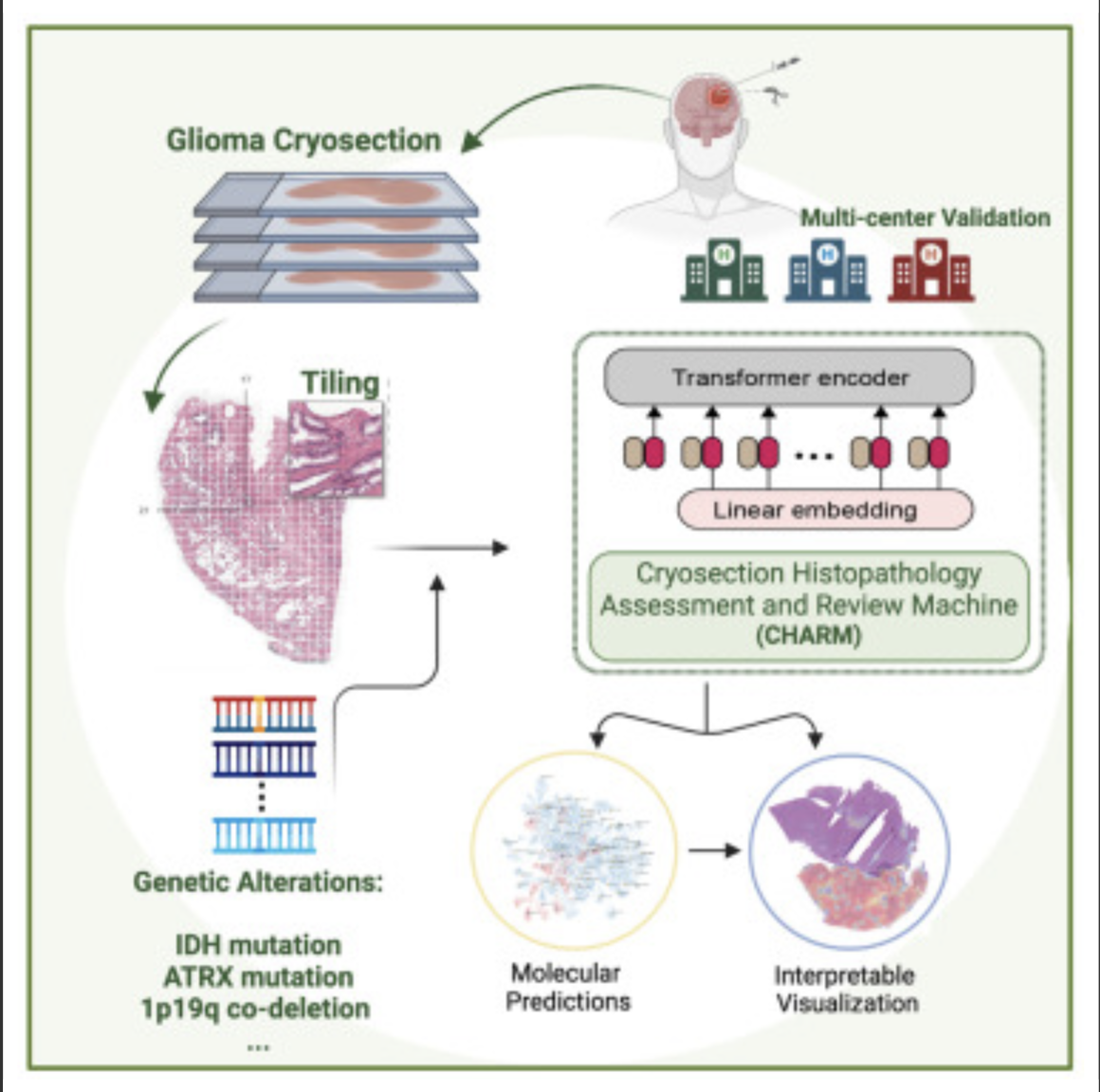
Kun-Hsing Yu and HMS colleagues used AI to rapidly determine a brain tumor’s molecular identity during surgery, propeling the development of precision oncology. The tool is CHARM (Cryosection Histopathology Assessment and Review Machine.) Currently, genetic sequencing takes days to weeks.  

Accurate molecular diagnosis during surgery can help a neurosurgeon decide how much brain tissue to remove. Removing too much when the tumor is less aggressive can affect a patient’s neurologic and cognitive function. Removing too little when the tumor is highly aggressive may leave behind malignant tissue that can grow and spread quickly. 

The technology will also allow the surgeon to determine if the patient can benefit from immediate treatment with drug-coated wafers placed directly into the brain at the time of the operation.

The standard intraoperative diagnostic approach used now involves taking brain tissue, freezing it, and examining it under a microscope. A major drawback is that freezing the tissue tends to alter the appearance of cells under a microscope and can interfere with the accuracy of clinical evaluation. Furthermore, the human eye, even when using potent microscopes, cannot reliably detect subtle genomic variations on a slide.

The new AI approach overcomes these challenges and could be particularly valuable in areas with limited access to technology to perform rapid cancer genetic sequencing.

Knowledge of a tumor’s molecular type provides insight about its aggressiveness, behavior, and likely response to various treatments, which can inform post-operative decisions.

The new tool enables during-surgery diagnoses aligned with the WHO classification system for diagnosing and grading the severity of gliomas, which calls for such diagnoses to be made based on a tumor’s genomic profile.

CHARM was developed using 2,334 brain tumor samples from 1,524 people with glioma from three different patient populations. When tested on a never-before-seen set of brain samples, the tool distinguished tumors with specific molecular mutations at 93 percent accuracy and successfully classified three major types of gliomas with distinct molecular features that carry different prognoses and respond differently to treatments.

It successfully captured visual characteristics of the tissue surrounding the malignant cells. It was capable of spotting telltale areas with greater cellular density and more cell death within samples, both of which signal more aggressive glioma types.

CHARM was also able to pinpoint clinically important molecular alterations in a subset of low-grade gliomas, a subtype of glioma that is less aggressive and therefore less likely to invade surrounding tissue. Each of these changes also signals different propensity for growth, spread, and treatment response.

It further connected the appearance of the cells — the shape of their nuclei, the presence of edema around the cells — with the molecular profile of the tumor. This means that the algorithm can pinpoint how a cell’s appearance relates to the molecular type of a tumor.

Accorging to Yu, this ability to assess the broader context around the image renders the model more accurate and closer to how a human pathologist would visually assess a tumor sample.

The researchers said that while the model was trained and tested on glioma samples, it could be successfully retrained to identify other brain cancer subtypes. 

Scientists have already designed AI models to profile other types of cancer — colon, lung, breast — but gliomas have remained particularly challenging due to their molecular complexity and huge variation in tumor cells’ shape and appearance.

The CHARM tool would have to be retrained periodically to reflect new disease classifications as they emerge from new knowledge. “Just like human clinicians who must engage in ongoing education and training, AI tools must keep up with the latest knowledge to remain at peak performance,” according to Yu.


Join ApplySci at MIT on September 18, 2023 for the 14th AI + Deep Tech Health + Neurotech conference.
Categories
AI Diabetes

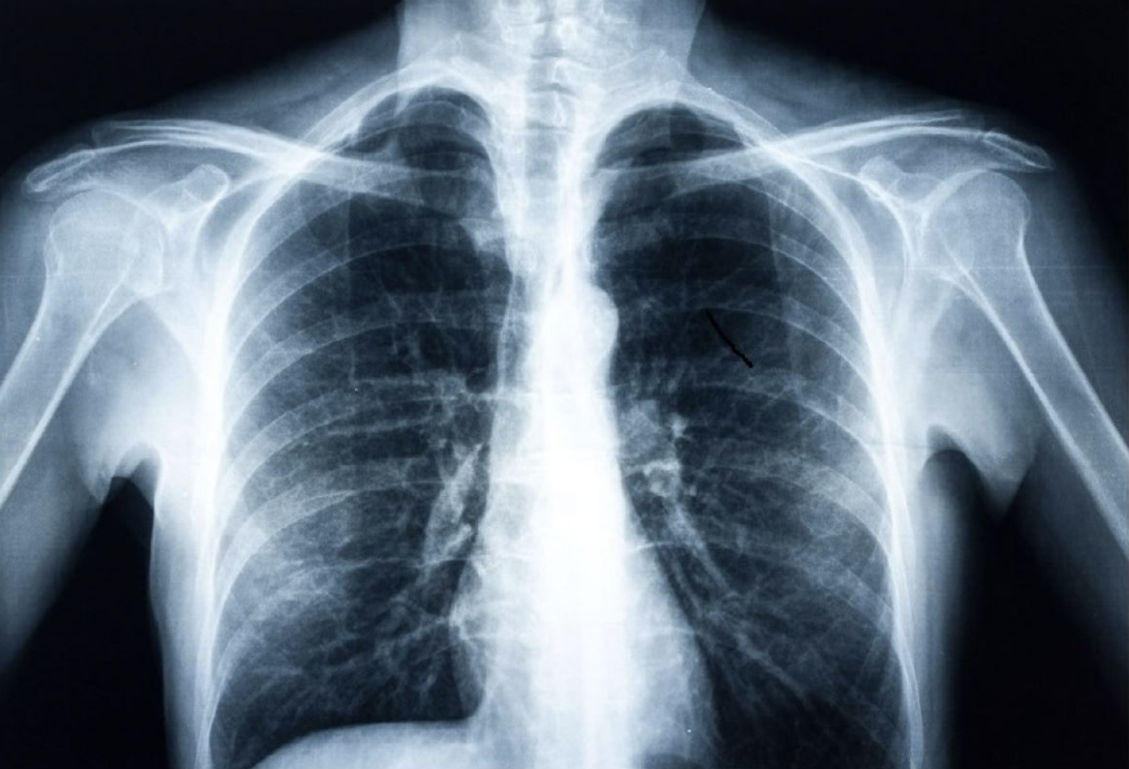
AI detects diabetes from fatty tissue in chest x rays

Judy Wawira Gichoya and Emory colleagues have developed an AI model that detects warning signs for diabetes in x rays collected during routine exams. The signs were also detected in patients who do not meet elevated risk guidelines.

Applying deep learning to images and electronic health record data, the model that successfully flagged elevated diabetes risk in a retrospective analysis, often years before patients were diagnosed.

Current guidelines suggest screening patients for type 2 diabetes if they are between 35 and 70 years old and have a body mass index (BMI) in the overweight to obese range. This strategy misses a significant number of cases, particularly in racial/ethnic minorities for whom BMI is a less effective predictor of diabetes risk.

Each year, millions of Americans receive chest x-rays for chest pain, difficulty breathing, injury or before surgeries. While radiologists are not looking for diabetes when they assess these x-rays, the images become part of a patient’s medical record and could be analyzed later for diabetes or other conditions.

The AI model was trained on more than 270,000 x-ray images from 160,000 patients, with deep learning determining the image features that best predicted a later diagnosis of diabetes. Because chest x-rays are not a common way to detect diabetes, the researchers also used explainable AI techniques to determine how and why the model made its determinations. The methods pointed to the location of fatty tissue as important for determining risk, a logic that aligns with recent medical findings that visceral fat in the upper body and abdomen is associated with type 2 diabetes, insulin resistance, hypertension and other conditions.

When the Emory team applied the model to a separate group of nearly 10,000 patients, they found the model predicted risk better than a simple model based on non-image clinical data alone.  

In some cases, the chest x-ray warned of high diabetes risk as early as three years before the patient eventually received a diagnosis. The model’s output also provides a numerical risk score that could potentially help clinicians customize the treatment approach for patients

The research team will now explore how to further validate the model and incorporate it into electronic health record systems so it can provide an alert to physicians to pursue traditional diabetes screening of patients flagged as high risk based on x-ray results. 

They’ll then turn to investigating how well chest x-rays can help diagnose other conditions, such as vascular disease, congestive heart failure, and chronic obstructive pulmonary disease.


Join ApplySci at MIT for the 14th AI + Deep Tech Health + Neurotech conference on September 18, 2023

Categories
AI Cancer

AI-based device increases ademona detection during colonoscopy

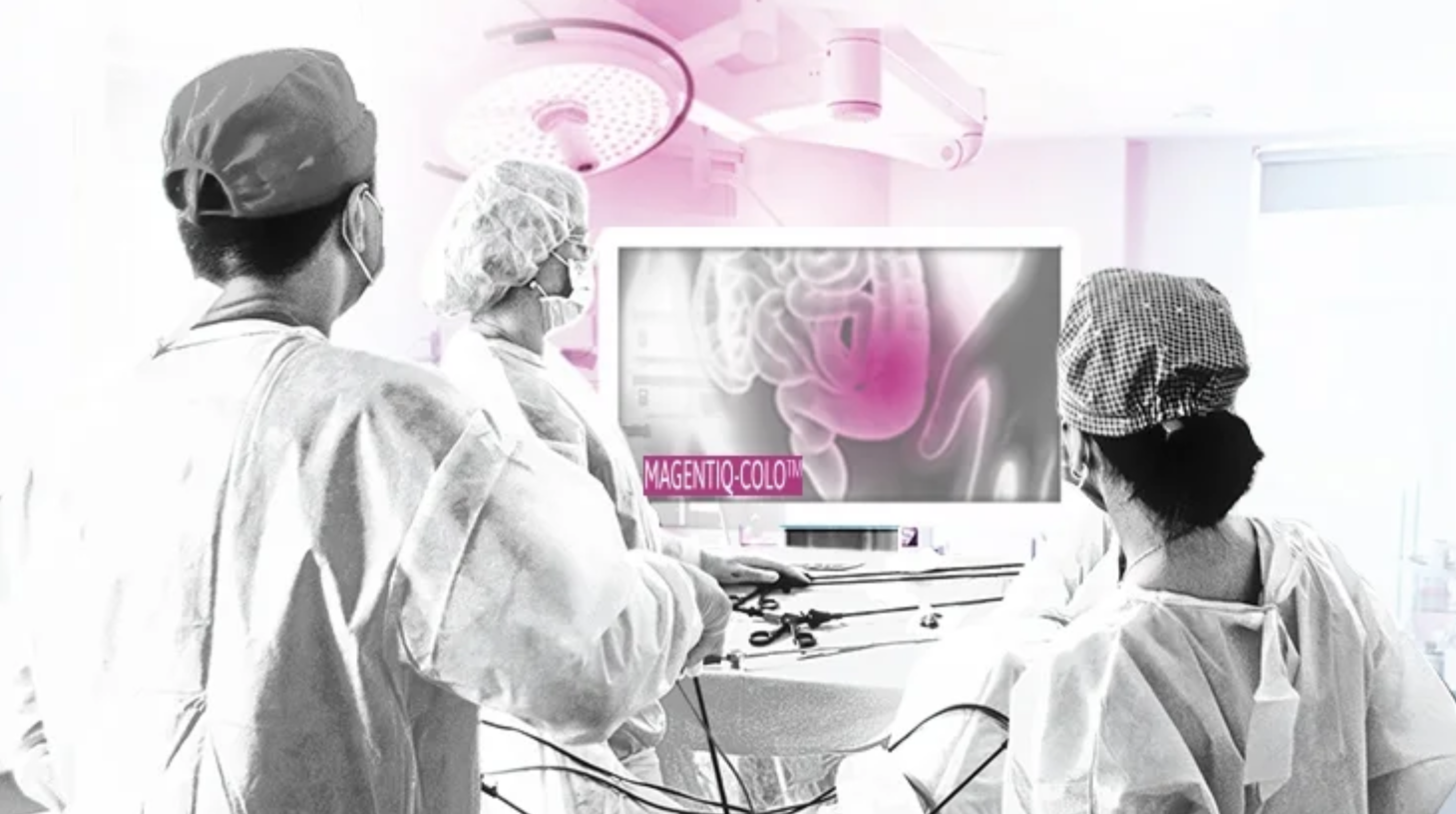
MAGENTIQ-COLO is an AI based FDA cleared colonoscopy device which offers a significant increase in Adenoma Detection Rate.

Current high rates of missed and undetected adenomas during colonoscopy means that even regularly screened patients are still at risk of developing colon cancer. A missed polyp can lead to interval cancer, which accounts for approximately 8% to 10% of all CRC in the U.S., translated to over 13,500 cancer cases that could be prevented every year with better detection.

A 2022 study of 950 patients at 10 hospitals showed that MAGENTIQ-COLO increasing ADR by 26% relatively, translating to a 21% decrease in CRC occurrence and a 35% decrease in patient mortality.


Join ApplySci at MIT on September 18, 2023 for AI + Deep Tech Health + Neurotech Boston

Categories
AI Brain Sensors

Biosensor detects misfiled proteins in Parkinson’s and Alzheimer’s disease

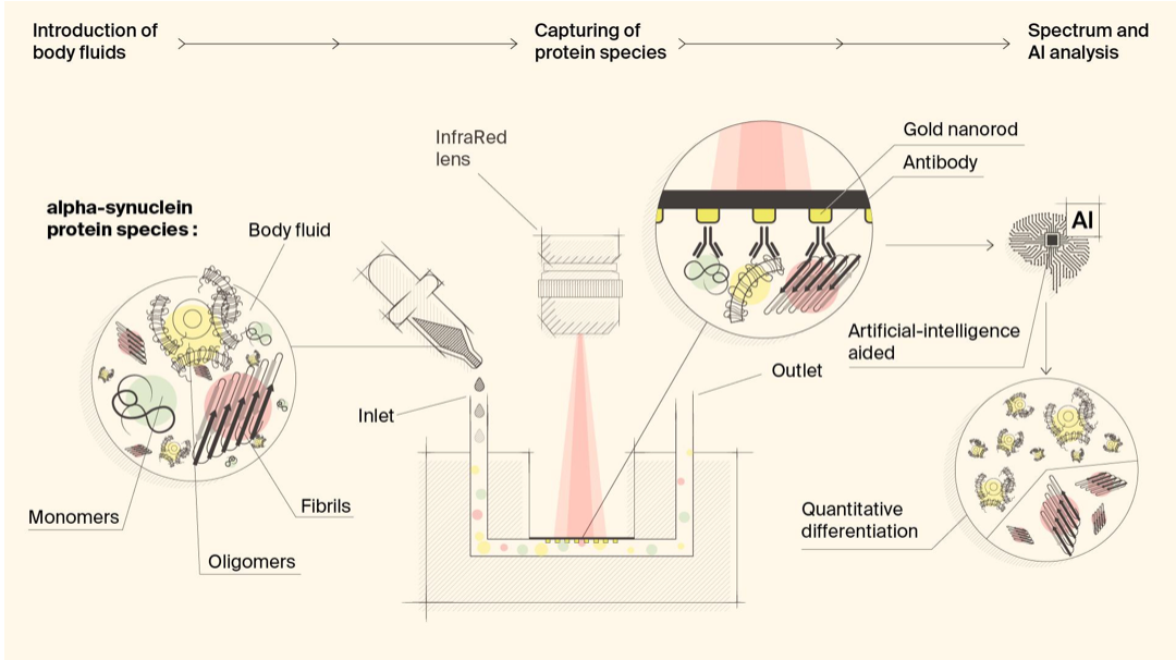
Hatice Altug, Hilal Lashue, and EPFL colleagues have developed ImmunoSEIRA, an AI-enhanced, biosensing tool for the detection of misfolded proteins linked to Parkinson’s and Alzheimer’s disease. The researchers also claim that neural networks can quantify disease stage and progression.

The technology holds promise for early detection, monitoring, and assessing treatment options.

Protein misfolding has been identified as a key event in disease progression. It is thought that healthy proteins misfold first into oligomers , and then into fibrils in later stages.

According to Lashuel, “unlike current biochemical approaches which rely on measuring the levels of these molecules, our approach is focused on detecting their abnormal structures. This technology also allows us to differentiate the levels of oligomers and fibrils.”

The sensor uses gold nanorod arrays with antibodies for specific protein detection, enabling real-time capture and structural analysis of target biomarkers from very small samples. Neural networks identify the presence of specific misfolded protein forms. Lashuel believes that “since the disease process is tightly associated with changes in protein structure, we believe that structural biomarkers, especially when integrated with other biochemical and neurodegeneration biomarkers, could pave the way for more precise diagnosis and monitoring of disease progression.”


Join ApplySci at MIT on September 18, 2023, for a scientist-led day of AI, Deep Tech Health , and Neurotech

Categories
AI Cancer

AI predicts pancreatic cancer

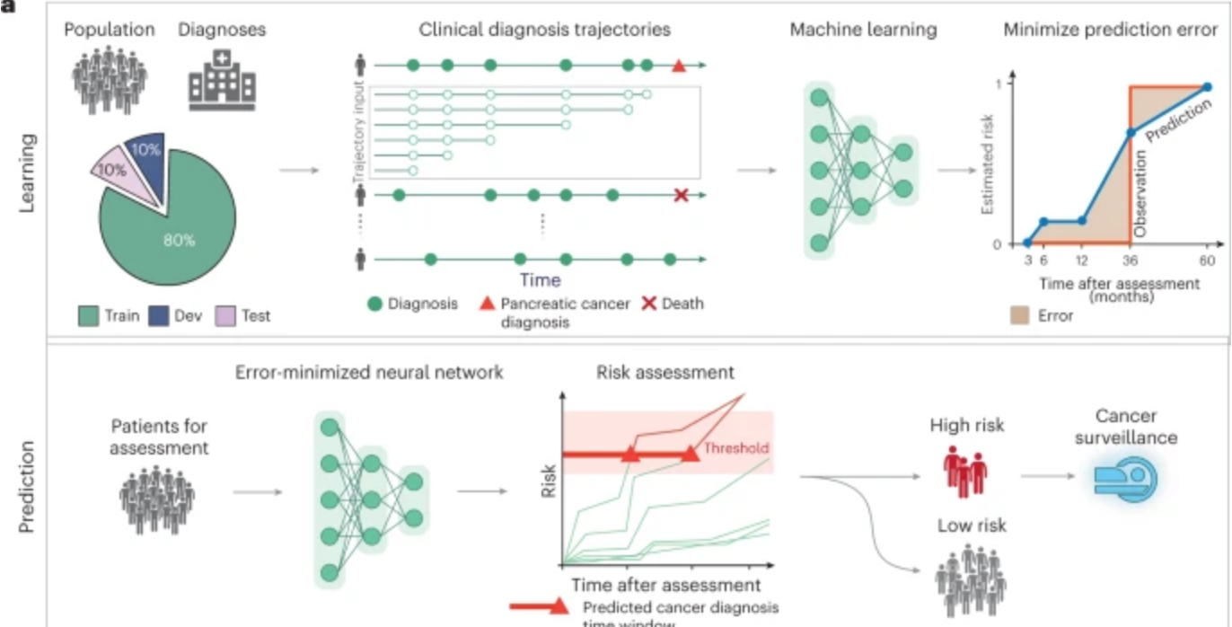
Harvard professor Chris Sander used clinical data from 6 million patients in Denmark’s national health system and 3 million in the U.S. VA system to train an AI model to predict the occurrence of pancreatic cancer within 3, 6, 12, and 36 months. This could allow wider screening for the aggressive disease, which is often discovered at a late stage, including in people with no known genetic risk.

The researchers believe that data from imaging, genetic, and wearable devices could further improve this tool, and that unrelated disease histories, including diabetes and substance abuse have already improved its accuracy.


Join ApplySci at MIT for the 14th AI+ Deep Tech + Neurotech conference on the future of healthcare on September 18, 2023

Categories
AI Brain

AI reconstructs viewed images

Yu TakagiShinji Nishimoto and Osaka University colleagues have published a  study which demonstrates that AI can read brain scans and re-create largely realistic versions of images a person has seen. Future applications could include enabling communication of people with paralysis, recording dreams, and understanding animal perception, among others.

Additional training was used on the existing text-to-image generative AI Stable Diffusion system, linking text descriptions about thousands of photos to brain patterns elicited when those photos were observed. Stable Diffusion was able to get more out of less training for each participant by incorporating photo captions into the algorithm.

The algorithm uses information gathered from regions of the brain involved in image perception, such as the occipital and temporal lobes. The system interpreted information from fMRI scans, detecting changes in blood flow to active brain regions. When people look at a photo, the temporal lobes register information about image contents (people, objects, or scenery), and the occipital lobe predominantly registers information about layout and perspective, such as the scale and position. This is recorded by the fMRI as it captures peaks in brain activity, and these patterns can then be reconverted into an imitation image using AI.

Categories
AI

Google AI detects tuberculosis

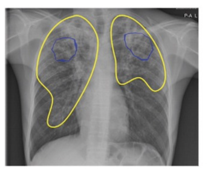
Google’s deep learning technology detected tuberculosis with similar accuracy to radiologists in a Radiology study.

165,174 chest radiographs from 22,284 patients in four countries were scanned. In detecting active tuberculosis, its sensitivity was higher (88 percent versus 75 percent) and its specificity was noninferior (79 percent versus 84 percent) compared to nine radiologists. Costs were reduced by 40 to 80 percent per tuberculosis-positive patient.

This may be able to facilitate tuberculosis screening in areas with limited radiologist resources, improving public health.


Join ApplySci at MIT for Deep Tech Health + Neurotech Boston on September 30th, with talks by:

Giovanni Traverso – Bob Langer – Emery Brown – George Church – Mary Lou Jepsen – Tom Oxley – Ellen Roche – Nathan Intrator – Bakul Patel – Ramita Tandon – Shaun Patel – Elizabeth Andukowich – Cris De Luca – Robert Garber – Jonathan Behr – Ann DeWitt

Categories
AI Brain Parkinson's

Neural Network assesses sleep patterns for passive Parkinson’s diagnosis

MIT’s Dina Katabi has developed a non-contact, neural network-based system to detect Parkinson’s disease while a person is sleeping.

By assessing nocturnal breathing patterns, the series of algorithms detects, and tracks the progression of, the disease — every night, at home.

A device in the bedroom emits radio signals, analyzes their reflections off the surrounding environment, and extracts breathing patterns, without bodily contact. The breathing signal is then fed to the neural network to assess Parkinson’s Disease in a passive manner.

Current diagnosis methods are invasive, expensive, and must be done at specialized centers, making frequent testing almost impossible.

Katabi said that a relationship between Parkinson’s and breathing was noted in 1817, motivating her to explore this form of detection, and that respiratory symptoms manifest years before motor symptoms.


Join ApplySci at MIT on September 30th for Deep Tech Health + Neurotech Boston

Categories
AI Cancer

AI catches breast cancer earlier, more often than traditional screening alone

The mammography screening paradigm has not changed since the 1960s.

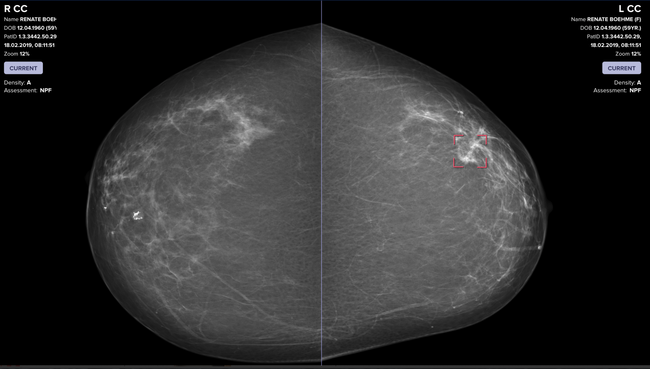
Breast screening AI company Vara, with Essen University and Memorial Sloan Kettering hospitals, published a study showing that radiologists assisted by AI are better able to screen for breast cancer. The hope is that AI systems could detect cancers that doctors miss, provide better care in remote areas, and allow radiologists more time to see more patients.

Two approaches were tested. In the first, AI analyzed mammograms. In the second, AI distinguished between normal and concerning scans. It refers the latter to a radiologist, who reviews them before seeing the AI’s assessment. Then the AI issues a warning if it detected cancer when the doctor did not. In the study, the AI examined old scans and compared its assessments with those of the radiologist who reviewed them.

Data from 367,000 mammograms—including radiologists’ notes, original assessments, and information on whether the patient ultimately had cancer— was analyzed to learn how to place these scans into one of three categories: “confident normal,” “not confident” (in which no prediction is given), and “confident cancer.” The conclusions from both approaches were then compared with the decisions real radiologists originally made on 82,851 mammograms sourced from screening centers that didn’t contribute scans used to train the AI.

The second approach—doctor and AI working together—was 2.6% better at detecting breast cancer than a doctor working alone, and raised fewer false alarms. It automatically set aside scans it classified as “confident normal,” which was 63% of all mammograms.


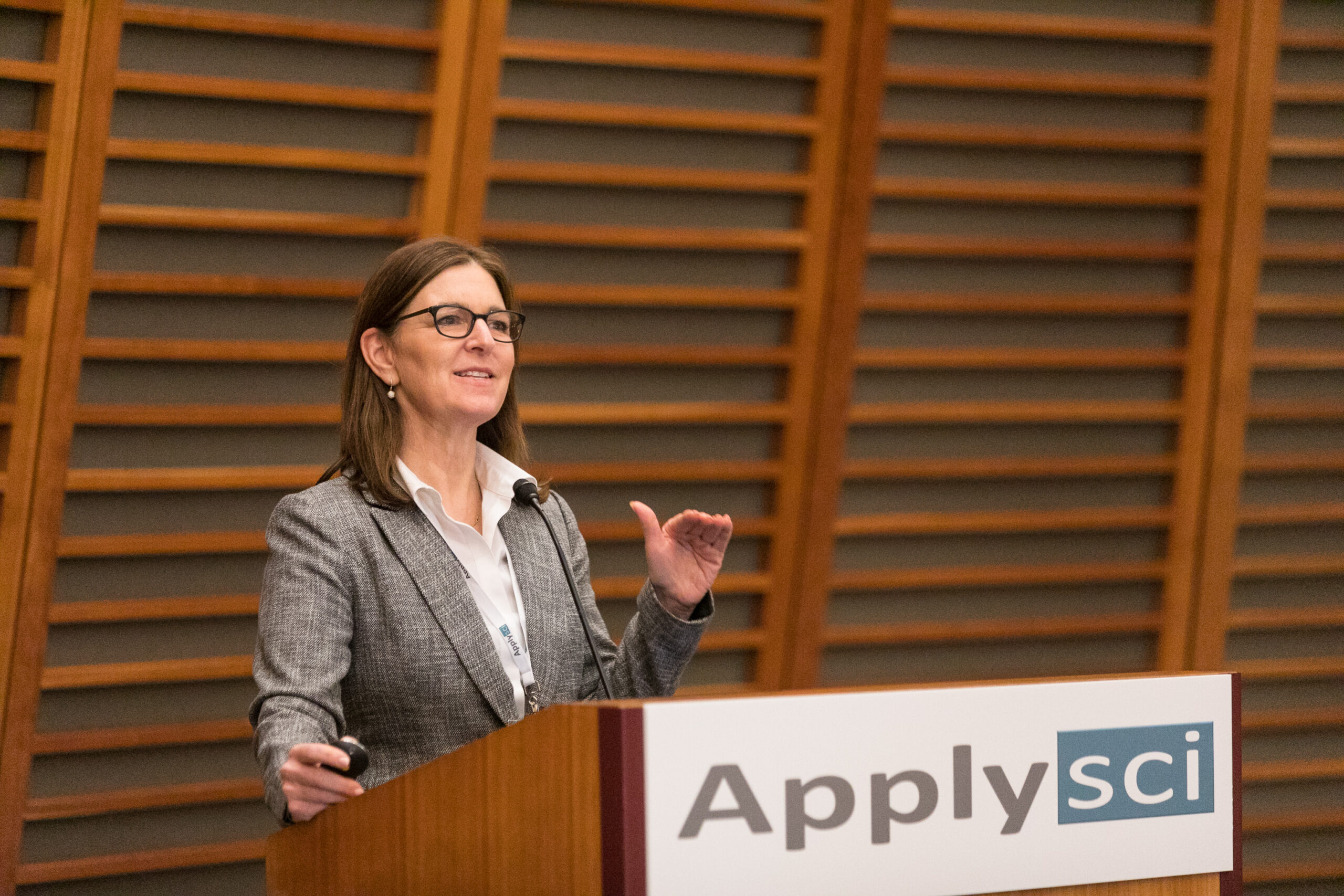
Clairity, an AI-driven precision breast cancer screening company, using Harvard professor Connie Lehman and colleagues’ decades of research in radiomics and applied AI, has developed a rigorous scientific approach to improve the accuracy of risk assessment. Prof Lehman discussed her approach at the 2019 ApplySci conference at Harvard Medical School.


Join ApplySci at MIT on September 30, 2022 for Deep Tech Health + Neurotech Boston featring Giovanni Traverso, MITBakul Patel, GoogleGeorge Church, Harvard Kerri Dugan, DARPAEllen Roche, MITNathan Intrator, NeurosteerEmery Brown, HarvardMary Lou Jepsen, OpenwaterShaun Patel, REACT NeuroElizabeth Andukowich, NIMHRamita Tandon, WalgreensCris de Luca, Sanofi VenturesTom Oxley, Synchron

Categories
AI Covid-19

AI detects COVID in chest x rays

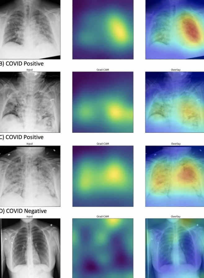
DeepCOVID-XR is a Northwestern University developed algorithm that automatically detects the signs of COVID-19 on a basic X-ray of the lungs. The system is able to detect COVID-19 in X-rays 10 times faster than thoracic radiologists and 1% to 6% more accurately.

The developers said the AI could be used to rapidly screen patients at hospital admission, and trigger protocols to help protect healthcare workers.

Accoring to Professor Aggelos Katsaggelos, the alorithm will not replace testing, but will enable the use of cheap, routine, safe x-rays to determine if a patient needs to be isolated.

In 17,000 X-ray images, the algorithm identified lungs which appeared patchy and hazy, as air sacs became inflamed and filled with fluid instead of air, which is common in COVID-19.

When put up against five experienced, fellowship-trained radiologists, DeepCOVID-XR was able to process a set of 300 test X-rays in about 18 minutes, compared to about two and a half to three and a half hours. The AI also delivered an accuracy rate of 82%, about on par with the group’s range of 76% to 81%.

“Radiologists are expensive and not always available,” Katsaggelos said. “X-rays are inexpensive and already a common element of routine care. This could potentially save money and time—especially because timing is so critical when working with COVID-19.”

Categories
AI Cancer

AI for (much) earlier breast cancer detection – Constance Lehman

Connie Lehman — Professor, Radiology, Harvard Medical School; Chief of Breast Imaging, radiology, Massachusetts General Hospital; Co-director of AVON Breast Center, Radiology, Massachusetts General Hospital; and Director, Breast Imaging Research Center, Radiology, Massachusetts General Hospital spoke at ApplySci’s recent conference at Harvard Medical School.

Click to listen to her talk on using AI to discover breast cancer 5 years earlier than currently possible.


Join ApplySci at the 13th Wearable Tech + Digital Health + Neurotech Silicon Valley conference on February 11-12, 2020 at Quadrus Sand Hill Road.  Speakers include:  Zhenan Bao, Stanford – Vinod Khosla, Khosla Ventures – Mark Chevillet, Facebook – Shahin Farshchi, Lux Capital – Carla Pugh, Stanford – Nathan Intrator, Tel Aviv University | Neurosteer – Wei Gao, Caltech – Sergiu Pasca, Stanford – Walter Greenleaf, Stanford – Sheng Xu, UC San Diego – Dror Ben-Zeev, University of Washington – Mikael Eliasson, Roche  – Unity Stoakes, StartUp Health

Categories
Conference

Wearable Tech + Digital Health + Neurotech Boston

Join ApplySci at the 12th Wearable Tech + Digital Health + Neurotech Boston conference on November 14, 2019 at Harvard Medical School featuring talks by Brad Ringeisen, DARPA – Joe Wang, UCSD – Carlos Pena, FDA  – George Church, Harvard – Diane Chan, MIT – Giovanni Traverso, Harvard | Brigham & Womens – Anupam Goel, UnitedHealthcare  – Nathan Intrator, Tel Aviv University | Neurosteer – Arto Nurmikko, Brown – Constance Lehman, Harvard | MGH – Mikael Eliasson, Roche – Nicola Neretti, Brown – R. Jacob  Vogelstein, Camden Partners – Yael Mandelblat-Cerf, Biogen