Categories
Brain Conference Parkinson's Ultrasound

Focused ultrasound thalamotomy in Parkinson’s Disease

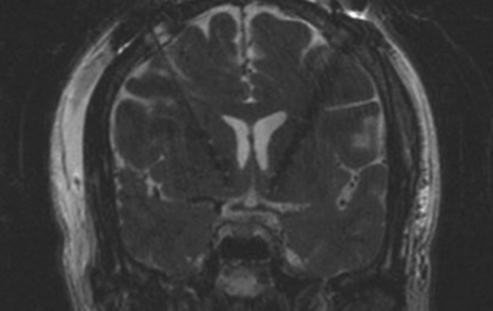
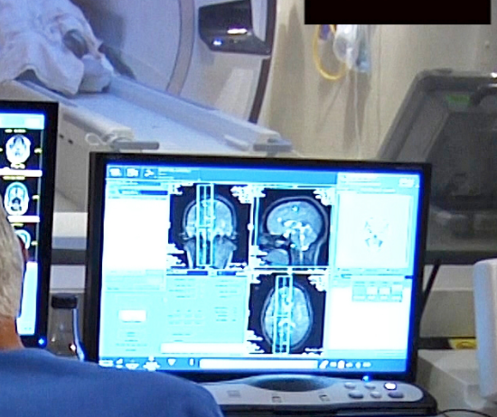
UVA’s Scott Sperling and Jeff Elias, who already used focused ultrasound to treat essential tremor, have just published the results of  a small study showing the efficacy of the technology in Parkinson’s Disease.

The sound waves were shown to interrupt brain circuits responsible for the uncontrollable shaking associated with the disease. The researchers claim that their study also offers “comprehensive evidence of safety” in its effect on mood, behavior and cognitive ability, which has not previously been studied.

According to Sperling, “In this study, we extended these initial results and showed that focused ultrasound thalamotomy is not only safe from a cognitive and mood perspective, but that patients who underwent surgery realized significant and sustained benefits in terms of functional disability and overall quality of life.”

27 adults with severe Parkinson’s tremor that had not responded to previous treatment were divided  into two groups. Twenty received the procedure, and a control group of seven (who were later offered the procedure) did not. Participants reported improved quality of life, including their ability to perform simple daily tasks, emotional wellbeing, and a lessened sense of stigma due to their tremor, at both three and twelve months.

The team found that mood and cognition, and the ability to go about daily life, ultimately had more effect on participants’ assessment of their overall quality of life than did remor severity or the amount of tremor improvement.

Cognitive decline was seen in some participants after the study, in that they were less able to name colors and think of and speak words. The cause of this was unclear, and must be investigated. The researchers suggested this could be a result of the natural progression of Parkinson’s.


Join ApplySci at the 10th Wearable Tech + Digital Health + Neurotech Silicon Valley conference on February 21-22 at Stanford University — Featuring:  Zhenan BaoChristof KochVinod KhoslaWalter Greenleaf – Nathan IntratorJohn MattisonDavid EaglemanUnity Stoakes Shahin Farshchi Emmanuel Mignot Michael Snyder Joe Wang – Josh Duyan – Aviad Hai Anne Andrews Tan Le

Categories
Brain Sensors

Wearable sensor monitors shunt function in hydrocephalus

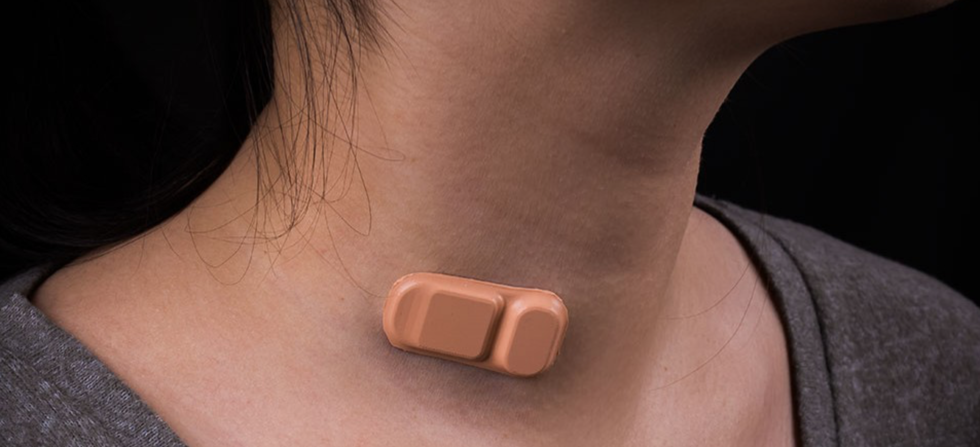
Northwestern’s John Rogers has created another minimal, flexible, wireless, adhesive wearable — this time to help hydrocephalus patients manage their condition.

The band-aid like sensor determines whether a shunt is working properly.

Shunts often fail.  When this happens, a patient can experience headaches, nausea and low energy, and must go to a hospital immediately.  However, a patient can have similar symptoms with a properly working shunt. The wearable determines, in five minutes, if the shunt is functioning, and if it is, a patient could avoid a hospital visit, CT, MRI, and potential surgery to determine the shunt’s functionality.

Click to view Northwestern University video


Join ApplySci at the 10th Wearable Tech + Digital Health + Neurotech Silicon Valley conference on February 21-22 at Stanford University — Featuring:  Zhenan BaoChristof KochVinod KhoslaWalter Greenleaf – Nathan IntratorJohn MattisonDavid EaglemanUnity Stoakes Shahin Farshchi Emmanuel Mignot Michael Snyder Joe Wang – Josh Duyan – Aviad Hai Anne Andrews Tan Le

Categories
Brain

Minimally invasive sensor detects electrical activity, optical signals in brain for MRI

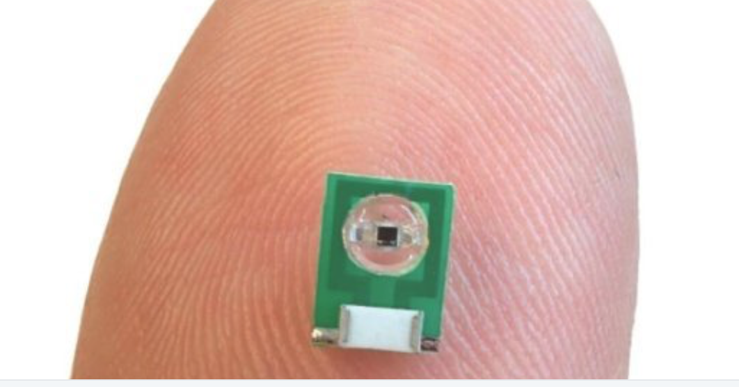
MIT’s Aviad Hai has developed a minimally invasive sensor to detect electrical activity or optical signals in the brain for MRI. No power source is needed, as radio signals that an external MRI scanner emits power the sensor.  It is implanted but does not require a wired connection to the brain. The researchers believe that it could also be adapted to measure glucose or other chemicals.

The team previously developed MRI sensors to detect calcium, serotonin and dopamine. The new sensor is meant to replace current electrical activity monitoring, which is extremely invasive, and can cause tissue damage.

Hai and colleagues shrank a radio antenna down to a few millimeters, so that it could be implanted directly into the brain to receive radio waves generated by water in the tissue.

The sensor is first tuned to the same frequency as the radio waves emitted by the hydrogen atoms. When an electromagnetic signal is detected, its tuning changes and  it no longer matches the hydrogen atom frequency. A weaker image then arises when the sensor is scanned by an external MRI machine.

In a study, the sensors were able to pick up electrical signals similar to those produced by action potentials or local field potentials.

Hai plans to further miniaturize the sensor, to enable multiple injections, to image light or electrical fields over a larger brain area.

Dr. Hai will discuss this work at ApplySci’s Wearable Tech + Digital Health + Neurotech Silicon Valley conference on February 21-22 at Stanford University


Join ApplySci at the 10th Wearable Tech + Digital Health + Neurotech Silicon Valley conference on February 21-22 at Stanford University — Featuring:  Zhenan BaoChristof KochVinod Khosla – Nathan IntratorJohn MattisonDavid EaglemanUnity Stoakes Shahin Farshchi – Emmanuel Mignot – Michael Snyder – Joe Wang – Josh Duyan – Aviad Hai

Categories
BCI Brain

Brain-to-brain communication interface

Rajesh Rao and University of Washington colleagues have developed BrainNet, a non-invasive direct brain-to-brain interface for multiple people.  The goal is a social network of human brains for problem solving. The interface combines EEG to record brain signals and TMS to deliver information to the brain, enabling 3 people to collaborate via direct brain-to-brain communication.

In a recent study, two of the three subjects were “Senders.” Their brain signals were decoded with real-time EEG analysis to extract decisions about whether to rotate a block in a Tetris-like game before it is dropped to fill a line. The Senders’ decisions were sent via the Internet to the brain of a third subject, the “Receiver.”  Decisions were delivered to the Receiver’s brain via magnetic stimulation of the occipital cortex. The Receiver integrated the information and made a decision, using an EEG interface, to either turn a block or keep it in the same position.  A second round of the game gave Senders another chance to validate and provide feedback to the Receiver’s action.


Join ApplySci at the 10th Wearable Tech + Digital Health + Neurotech Silicon Valley conference on February 21-22 at Stanford University — Featuring:  Zhenan BaoChristof KochVinod Khosla – Nathan IntratorJohn MattisonDavid EaglemanUnity Stoakes Shahin Farshchi

Categories
Brain Parkinson's

Implanted sensors track dopamine for a year

Helen Schwerdt, Ann Graybiel, Michael Cima, Bob Langer, and MIT colleagues have developed and implantable sensor that can measure dopamine in the brain of rodents for more than one year.  They believe that this can inform the treatment and understanding of Parkinson’s and other brain diseases.

According to Graybiel, “Despite all that is known about dopamine as a crucial signaling molecule in the brain, implicated in neurologic and neuropsychiatric conditions as well as our abilty to learn, it has been impossible to monitor changes in the online release of dopamine over time periods long enough to relate these to clinical conditions.”

The sensors arenearly invisible to the immune system, avoiding scar tissue that would impede accuracy. After  implantation, populations of microglia  and astrocytes were the same as those in brain tissue that did not have the probes.

In a recent animal  study, three to five sensors per were implanted 5 millimeters deep in the striatum. Readings were taken every few weeks, after dopamine release was stimulated in the brainstem, traveling to the striatum. Measurements remained consistent for up to 393 days.

If developed for use in humans, these sensors could be useful for monitoring Parkinson’s patients who receive deep brain stimulation.


Join ApplySci at the 9th Wearable Tech + Digital Health + Neurotech Boston conference on September 24, 2018 at the MIT Media Lab.  Speakers include:  Rudy Tanzi – Mary Lou Jepsen – George ChurchRoz PicardNathan IntratorKeith JohnsonJohn MattisonRoozbeh GhaffariPoppy Crum – Phillip Alvelda Marom Bikson – Ed Simcox – Sean Lane

Categories
Brain Virtual Reality

VR + motion capture to study movement, sensory processing, in autism, AD, TBI

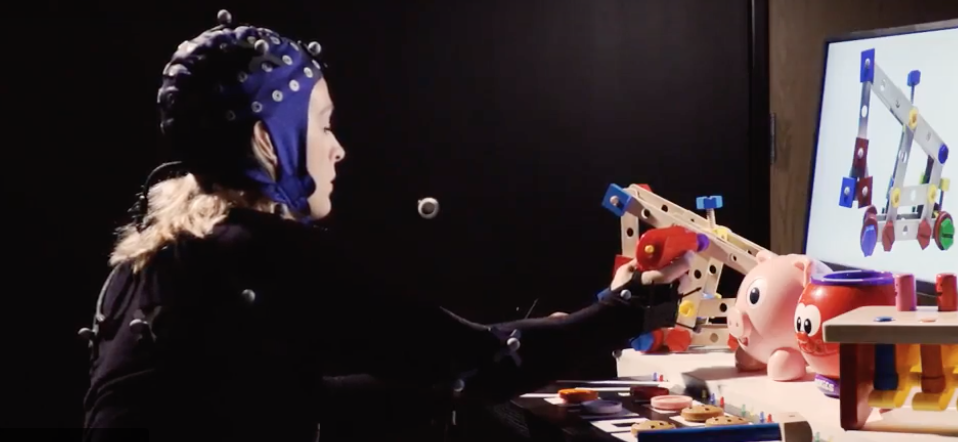
MoBi, developed by John Foxe at the University of Rochester, combines VR, EEG, and motion capture sensors to study movement difficulties associated with neurological disorders.

According to Foxe, “The MoBI system allows us to get people walking, using their senses, and solving the types of tasks you face every day, all the while measuring brain activity and tracking how the processes associated with cognition and movement interact.”

Motion sensor and EEG data, collected while a subject is walking in a virtual environment, are synchronized, allowing researchers to track which areas of the brain are being activated when walking or performing task. Brain response while moving, performing tasks, or doing both at the same time, is analyzed.

This technique could potentially guide treatment in Autism, dementia, and TBI, characterized by difficulty in processing sensory information from multiple sources and an abnormal gait.

Click to view University of Rochester video


Join ApplySci at the 9th Wearable Tech + Digital Health + Neurotech Boston conference on September 24, 2018 at the MIT Media Lab.  Speakers include:  Rudy Tanzi – Mary Lou Jepsen – George ChurchRoz PicardNathan IntratorKeith JohnsonJohn MattisonRoozbeh GhaffariPoppy Crum – Phillip Alvelda Marom Bikson – Ed Simcox – Sean Lane

PREFERRED REGISTRATION AVAILABLE THROUGH TODAY, SEPTEMBER 7TH

Categories
Brain

Brain imaging to detect suicidal thoughts

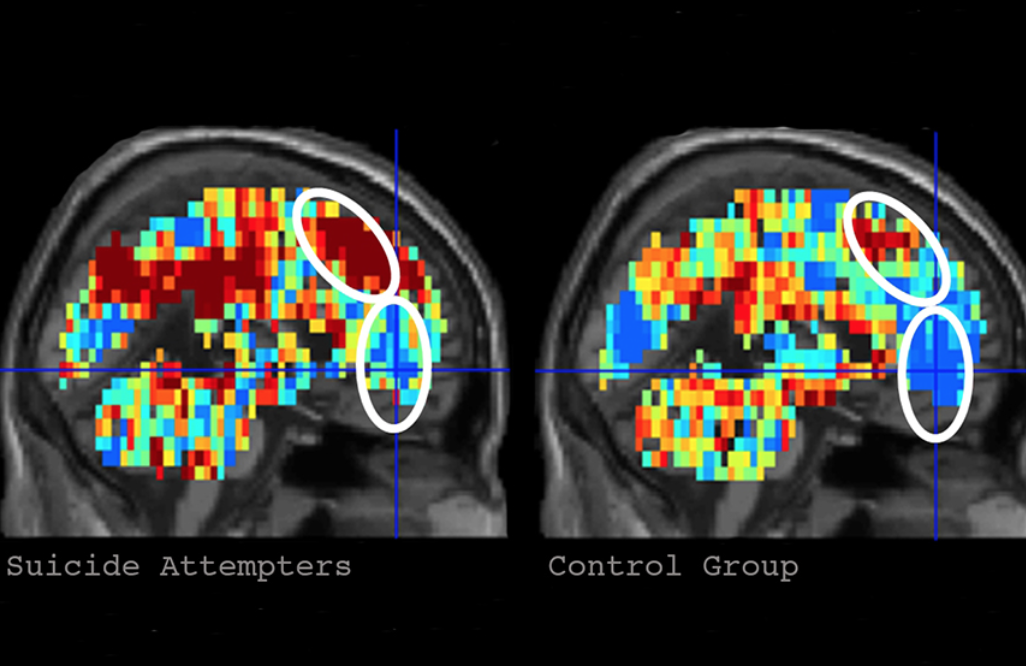
Last year, Carnegie Mellon professor Marcel Just and Pitt professor David Brent used brain imagining to identify suicidal thoughts.

Supported by the NIMH, they are now working to establish reliable neurocognitive markers of suicidal ideation and attempt. They will examine the differences in brain activation patterns between suicidal and non-suicidal young adults as they think about words related to suicide — such as positive and negative concepts — and use machine learning to identify neural signatures of suicidal ideation and behavior.

According to Just,  “We were previously able to obtain consistent neural signatures to determine whether someone was thinking about objects like a banana or a hammer by examining their fMRI brain activation patterns. But now we are able to tell whether someone is thinking about ‘trouble’ or ‘death’ in an unusual way. The alterations in the signatures of these concepts are the ‘neurocognitive thought markers’ that our machine learning program looks for.”


Join ApplySci at the 9th Wearable Tech + Digital Health + Neurotech Boston conference on September 24, 2018 at the MIT Media Lab.  Speakers include:  Rudy Tanzi – Mary Lou Jepsen – George ChurchRoz PicardNathan IntratorKeith JohnsonJohn MattisonRoozbeh GhaffariPoppy Crum – Phillip Alvelda Marom Bikson – Ed Simcox – Sean Lane

PREFERRED REGISTRATION AVAILABLE THROUGH FRIDAY, SEPTEMBER 7TH

Categories
AI Brain

AI predicts response to antipsychotic drugs, could distinguish between disorders

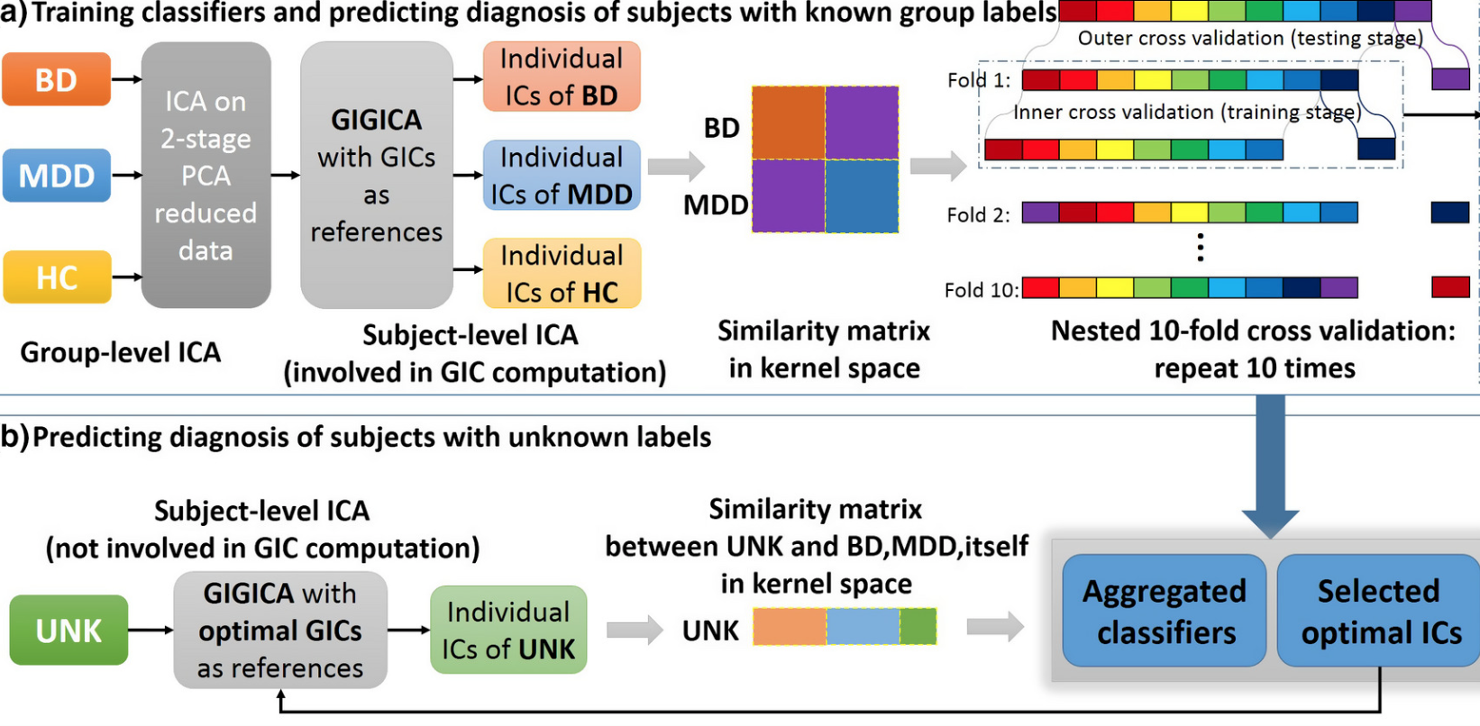
Lawson Health Research Institute, Mind Research Network and Brainnetome Center researchers have developed an algorithm that analyzes brain scans to classify illness in patients with complex mood disorders and help predict their response to medication.

A recent study analyzed and compared fMRI scans of those with MDD, bipolar I,  and no history of mental illness, and found that each group’s brain networks differed, including regions in the default mode network and thalamus.

When tested against participants with a known MDD or Bipolar I diagnosis, the algorithm correctly classified illness with 92.4 per cent accuracy.

The team also imaged the brains of 12 complex mood disorder patients with out a clear diagnosis, to predict diagnosis and examine medication response.

The researchers hypothesized that participants classified by the algorithm as having MDD would respond to antidepressants while those classified as having bipolar I would respond to mood stabilizers. When tested with the complex patients, 11 out of 12 responded to the medication predicted by the algorithm.

According to lead researcher Elizabeth Osuch:: “This study takes a major step towards finding a biomarker of medication response in emerging adults with complex mood disorders. It also suggests that we may one day have an objective measure of psychiatric illness through brain imaging that would make diagnosis faster, more effective and more consistent across health care providers.”


Join ApplySci at the 9th Wearable Tech + Digital Health + Neurotech Boston conference on September 24, 2018 at the MIT Media Lab.  Speakers include:  Rudy Tanzi – Mary Lou Jepsen – George ChurchRoz PicardNathan IntratorKeith JohnsonJuan EnriquezJohn MattisonRoozbeh GhaffariPoppy Crum – Phillip Alvelda Marom Bikson – Ed Simcox – Sean Lane

Categories
Brain

Invasive deep brain stimulation for alcoholism?

Stanford’s Casey Halpern and Allen Ho have used deep brain stimulation to target nucleus accumbens, thought to reduce impulsive behavior, to combat alcoholism in animal and pilot human studies.

DBS is used in severe Parkinson’s disease and is not approved by the FDA for addiction. Infection and other complications are risks of this invasive surgery.

ApplySci hopes that strides in behavioral therapy, including Alcoholics Anonymous, will continue to improve outcomes in addicted individuals, diminishing the need for invasive procedures.

The Stanford study was published in Neurosurgical Focus.


Join ApplySci at the 9th Wearable Tech + Digital Health + Neurotech Boston conference on September 24, 2018 at the MIT Media Lab.  Speakers include:  Rudy Tanzi – Mary Lou Jepsen – George ChurchRoz PicardNathan IntratorKeith JohnsonJuan EnriquezJohn MattisonRoozbeh GhaffariPoppy Crum – Phillip Alvelda Marom Bikson – Ed Simcox – Sean Lane

Categories
AI Brain Cancer

AI – optimized glioblastoma chemotherapy

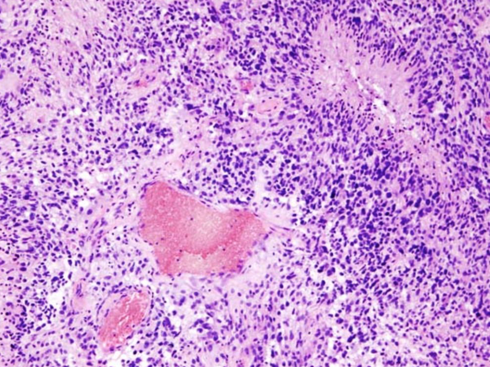
Pratik Shah, Gregory Yauney,  and MIT Media Lab researchers have developed an AI  model that could make glioblastoma chemotherapy regimens less toxic but still effective. It analyzes current regimens and iteratively adjusts doses to optimize treatment with the lowest possible potency and frequency toreduce tumor sizes.

In simulated trials of 50 patients, the machine-learning model designed treatment cycles that reduced the potency to a quarter or half of the doses It often skipped administration, which were then scheduled twice a year instead of monthly.

Reinforced learning was used to teach the model to favor certain behavior that lead to a desired outcome.  A combination of  temozolomide and procarbazine, lomustine, and vincristine, administered over weeks or months, were studied.

As the model explored the regimen, at each planned dosing interval it decided on actions. It either initiated or withheld a dose. If it administered, it then decided if the entire dose, or a portion, was necessary. It pinged another clinical model with each action to see if the the mean tumor diameter shrunk.

When full doses were given, the model was penalized, so it instead chose fewer, smaller doses. According to Shah, harmful actions were reduced to get to the desired outcome.

The J Crain Venter Institute’s Nicholas Schork said that the model offers a major improvement over the conventional “eye-balling” method of administering doses, observing how patients respond, and adjusting accordingly.


Join ApplySci at the 9th Wearable Tech + Digital Health + Neurotech Boston conference on September 24, 2018 at the MIT Media Lab.  Speakers include:  Rudy Tanzi – Mary Lou Jepsen – George ChurchRoz PicardNathan IntratorKeith JohnsonJuan EnriquezJohn MattisonRoozbeh GhaffariPoppy Crum – Phillip Alvelda Marom Bikson – Ed Simcox – Sean Lane

Categories
Brain

Sensor could continuously monitor brain aneurysm treatment

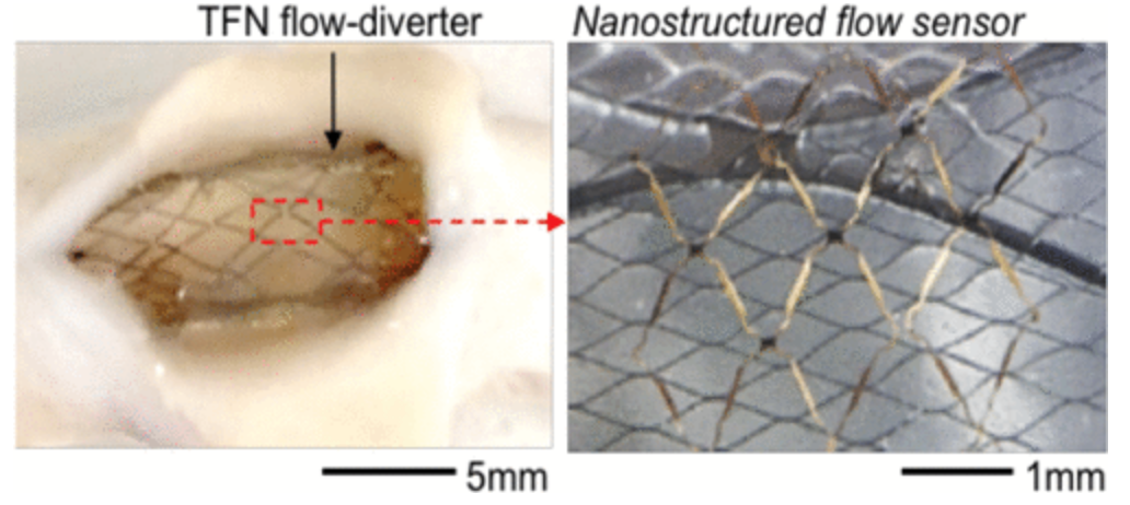
Georgia Tech’s Woon-Hong Yeo has  developed a proof of concept, flexible, stretchable sensor that can continuously monitor hemodynamics when integrated with a stent like flow diverter after a brain aneurysm. Blood flow is measured using  capacitance changes.

According to Pittsburgh professor Youngjae Chun, who collaborated with Yeo, “We have developed a highly stretchable, hyper-elastic flow diverter using a highly-porous thin film nitinol,” Chun explained. “None of the existing flow diverters, however, provide quantitative, real-time monitoring of hemodynamics within the sac of cerebral aneurysm. Through the collaboration with Dr. Yeo’s group at Georgia Tech, we have developed a smart flow-diverter system that can actively monitor the flow alterations during and after surgery.”

The goal is a batteryless, wireless device that is extremely stretchable and flexible that can be miniaturized enough to be routed through the tiny and complex blood vessels of the brain and then deployed without damage  According to Yeo, “It’s a very challenging to insert such electronic system into the brain’s narrow and contoured blood vessels.”

The sensor uses a micro-membrane made of two metal layers surrounding a dielectric material, and wraps around the flow diverter. The device is a few hundred nanometers thick, and is produced using nanofabrication and material transfer printing techniques, encapsulated in a soft elastomeric material.

“The membrane is deflected by the flow through the diverter, and depending on the strength of the flow, the velocity difference, the amount of deflection changes,” Yeo explained. “We measure the amount of deflection based on the capacitance change, because the capacitance is inversely proportional to the distance between two metal layers.”

Because the brain’s blood vessels are so small, the flow diverters can be no more than five to ten millimeters long and a few millimeters in diameter. That rules out the use of conventional sensors with rigid and bulky electronic circuits.

“Putting functional materials and circuits into something that size is pretty much impossible right now,” Yeo said. “What we are doing is very challenging based on conventional materials and design strategies.”

The researchers tested three materials for their sensors: gold, magnesium and the nickel-titanium alloy known as nitinol. All can be safely used in the body, but magnesium offers the potential to be dissolved into the bloodstream after it is no longer needed.

The proof-of-principle sensor was connected to a guide wire in the in vitro testing, but Yeo and his colleagues are now working on a wireless version that could be implanted in a living animal model. While implantable sensors are being used clinically to monitor abdominal blood vessels, application in the brain creates significant challenges.

“The sensor has to be completely compressed for placement, so it must be capable of stretching 300 or 400 percent,” said Yeo. “The sensor structure has to be able to endure that kind of handling while being conformable and bending to fit inside the blood vessel.”


Join ApplySci at the 9th Wearable Tech + Digital Health + Neurotech Boston conference on September 24, 2018 at the MIT Media Lab.  Speakers include:  Rudy Tanzi – Mary Lou Jepsen – George ChurchRoz PicardNathan IntratorKeith JohnsonJuan EnriquezJohn MattisonRoozbeh GhaffariPoppy Crum – Phillip Alvelda Marom Bikson – Ed Simcox – Sean Lane

Categories
Brain Heart Virtual Reality

David Axelrod: VR in healthcare & the Stanford Virtual Heart | ApplySci @ Stanford

David Axelrod discussed VR-based learning in healthcare, and the Stanford Virtual Heart, at ApplySci’s recent Wearable Tech + Digital Health + Neurotech conference at Stanford;


Join ApplySci at the 9th Wearable Tech + Digital Health + Neurotech Boston conference on September 24, 2018 at the MIT Media Lab.  Speakers include:  Rudy Tanzi – Mary Lou Jepsen – George ChurchRoz PicardNathan IntratorKeith JohnsonJuan EnriquezJohn MattisonRoozbeh GhaffariPoppy Crum – Phillip Alvelda Marom Bikson

REGISTRATION RATES INCREASE JULY 6th