Categories
Heart Monitoring Respiratory

Phone tracks health with out wearable sensors

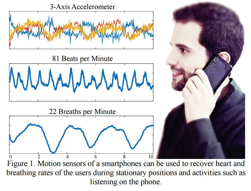
Javier Hernandez Rivera of Rosalind Picard‘s Affective Computing Group at MIT is developing a health monitoring phone that does not require a wearable.  BioPhone derives biological signals from a phone’s accelerometer, which the team says captures small body movements that result from one’s heart beating and chest rising and falling.

Hernandez said that BioPhone is meant to gather data during still moments, simplifying the capture of small vibrations without having to account for many body movements.   He believes that this can detect stress, which could trigger the phone to provide breathing exercises, or notify a loved one to call.

12 subjects sat, stood, and lied down, before and after pedaling a bike, with a smartphone in their pocket.  To compare results,  they wore sensors to capture heart and breathing rates. Heart rates reported by smartphone data alone were off by 1 beat per minute, and breathing rates were off by 1/4 of a breath per minute.

MIT Technology Review reported that the findings were questioned by a U of Alabama mobile health expert, who believes  that results will be affected by signal noise from inadvertent motions.

WEARABLE TECH + DIGITAL HEALTH SAN FRANCISCO – APRIL 5, 2016 @ THE MISSION BAY CONFERENCE CENTER

NEUROTECH SAN FRANCISCO – APRIL 6, 2016 @ THE MISSION BAY CONFERENCE CENTER
Categories
Heart Wearables

Chip uses ECG to monitor heart, improving wearable accuracy

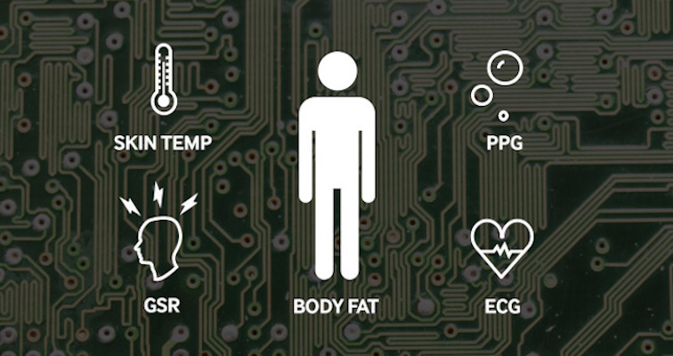
Samsung’s  Bio Processor uses ECG  to continuously measure the electric activity of the heart, improving its accuracy.  It must be worn on the skin near the heart, and includes built-in flash memory and Bluetooth for connecting to one’s phone.  The chip can also monitor PPG, ECG, skin temperature, GSR and body fat. The company describes this as a breakthrough in wearable accuracy.  While not yet available to consumers, it will be used in wearables for health, fitness, or biometric identification purposes.

WEARABLE TECH + DIGITAL HEALTH SAN FRANCISCO – APRIL 5, 2016 @ THE MISSION BAY CONFERENCE CENTER

NEUROTECH SAN FRANCISCO – APRIL 6, 2016 @ THE MISSION BAY CONFERENCE CENTER
Categories
3-D Printing Heart

Toward a 3D printed heart

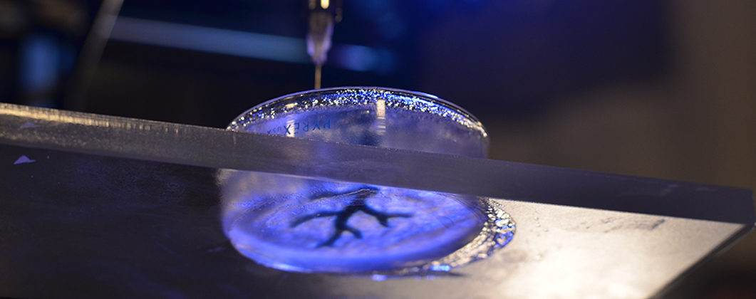
Carnegie Mellon‘s Adam Feinberg is developing 3D printing techniques that could in the future be used to repair the heart.  This work is aimed at alternative solutions for the 4,000 Americans currently waiting to receive a heart transplant.

Feinberg described his progress:  “We’ve been able to take MRI images of coronary arteries and 3-D images of embryonic hearts and 3-D bioprint them with unprecedented resolution and quality out of very soft materials like collagens, alginates and fibrins.”

The next step is to incorporate real heart cells into these 3-D printed tissue structures, providing a scaffold to help form contractile muscle.

Click to view Carnegie Mellon video.

WEARABLE TECH + DIGITAL HEALTH SAN FRANCISCO – APRIL 5, 2016 @ THE MISSION BAY CONFERENCE CENTER

NEUROTECH SAN FRANCISCO – APRIL 6, 2016 @ THE MISSION BAY CONFERENCE CENTER
Categories
Heart

Optogenetics used to regulate heartbeat

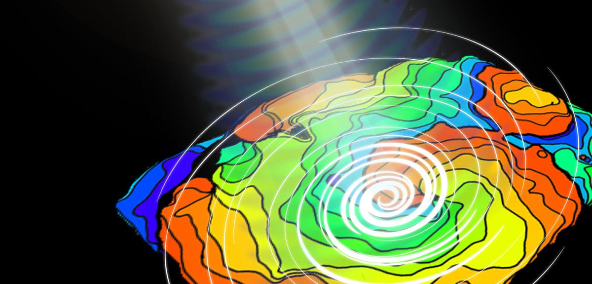
Oxford’s Gil Bub and and Stony Brooks’ Emilia Entcheva have used optogenetics, a method used to control neurons, to regulate heartbeat.

Arrhythmia patients currently use pacemakers or drugs to control heart rhythm.   These approaches can stop or start waves, but cannot provide fine control over wave speed and direction.

In the Bub and Entcheva study, a protein called channelrhodopsin was delivered to heart cells via gene therapy.  This made them light-responsive. Using a computer-powered light projector, the researchers controlled  the speed and direction of the cardiac waves, and the orientation of spirals, in real time.  This has never before been shown in a living system.

Click to view University of Oxford video.

WEARABLE TECH + DIGITAL HEALTH SAN FRANCISCO – APRIL 5, 2016 @ THE MISSION BAY CONFERENCE CENTER

NEUROTECH SAN FRANCISCO – APRIL 6, 2016 @ THE MISSION BAY CONFERENCE CENTER
Categories
Heart

Noninvasive 3D scan identifies coronary artery blockages

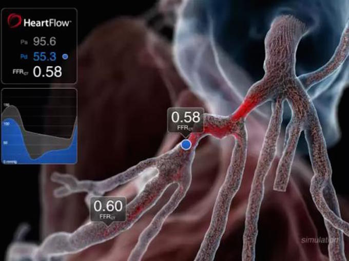
Fractional flow reserve computed tomography is a high-definition 3D scanner used to identify blocked arteries around the heart.  The technology, developed by Heartflow, eliminates the risk of diagnostic methods where vessels are probed.

The risk of death from an angiogram is one in 1,000, and can be caused by a rupture of an artery, or a stroke or heart attack caused by fatty plaque breaking off during the test.

FFRCT creates a detailed computer model of the heart from a CT image. It then (noninvasively) calculates the extent of blockages in the coronary arteries and whether they are restricting the flow of blood.

WEARABLE TECH + DIGITAL HEALTH SAN FRANCISCO – 4/5/16 @ MISSION BAY CONFERENCE CENTER
NEUROTECH SAN FRANCISCO – 4/6/16 @ MISSION BAY CONFERENCE CENTER
Categories
Heart

Device detects heart attacks with one drop of blood

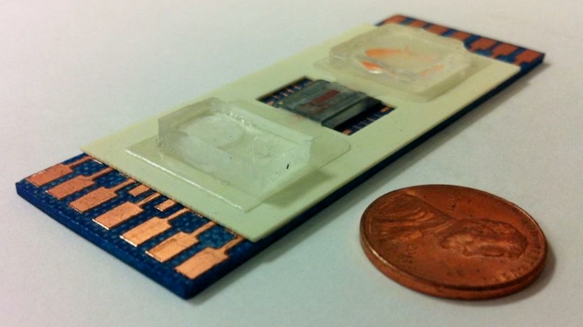
UCLA’s Chi On Chui has developed a device that brings lab quality bio-molecular assessments to point-of-care settings, such as clinics, ambulances or homes.

SELFA (Semiconductor Electronic Label-Free Assay) could reduce emergency room time for heart attack patients by hours. Similar to a diabetic glucose sensor, SELFA uses a single drop of blood, taken wherever symptoms occur, to determine which patients do not require further medical care.

According to Chui: “Eighty-five percent of patients who present with symptoms of acute coronary syndrome in emergency rooms across the U.S. and Europe — some 12.75 million per year — have actually not had heart attacks after a standard test comes back, hours later, from the lab.”

WEARABLE TECH + DIGITAL HEALTH SAN FRANCISCO – APRIL 5, 2016 – REGISTER HERE

NEUROTECH SAN FRANCISCO – APRIL 6, 2016 – REGISTER HERE

Categories
3-D Printing Heart

Faster, personalized, 3D printed heart models for surgery planning

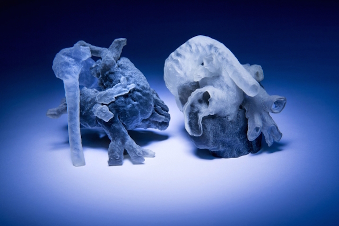
MIT and Boston Children’s Hospital researchers are converting heart MRI scans into 3D printed physical models,  for surgical planning,  in 3-4 hours.  Previously, the process took 10 hours. The project, which limits human input to increase accuracy, is led by Professor Polina Golland.  Physicist Medhi Moghari enhanced the precision of the MRI, decreasing the dependence on generic models, and enabling the the team to create the algorithm and print the model in the shorter time frame.

The algorithm examines patches of unsegmented cross sections and looks for similar features in the nearest segmented cross sections. Golland believes that its performance might be improved if it also examined patches that ran obliquely across several cross sections, which will be the next phase of research.

Categories
Heart Monitoring Respiratory

Piezoelectric sensor car seat monitors respiration, heart rate

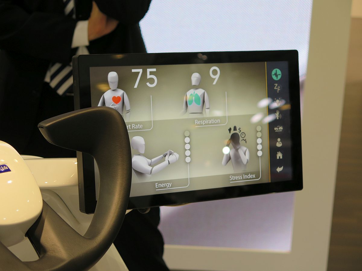
Faurecia‘s “Active Welness” car seat monitors respiration and heart rate with embedded piezoelectric sensors.  The goal is to detect driver stress or alertness.  When low energy is detected, the seat responds with specific massage patterns and air flow through the ventilation system.  The non-contact sensors were developed by Hoana Medical.  Combined with advanced algorithms and signal processing, Faurecia claims that they can accommodate noise and vibration from the moving vehicle without compromising effectiveness.

Categories
Heart Monitoring

Simple, rapid “tricorder” vital sign monitoring

Johns Hopkins researchers  have developed a “tricorder” that quickly picks up vital signs from a patient’s lips and fingertip.

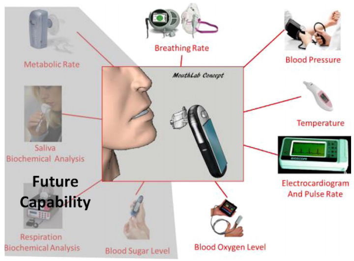
MouthLab could replace bulky monitors and gather more data during  an ambulance, emergency room, doctor’s office or home assessment.

Heart rate, blood pressure, temperature, breathing rate, blood oxygen and a basic ECG are measured.   Early signs of emergencies, such as heart attacks, could be detected.

Monitoring by mouth will allow future versions to detect chemical cues in blood, saliva and breath that act as markers for health conditions.   According to Professor Gene Fridman: “We envision the detection of a wide range of disorders,from blood glucose levels for diabetics, to kidney failure, to oral, lung and breast cancers.”

Categories
Heart

Cheap, non-invasive, mobile cardiovascular disease screening

An IMEC led consortium is developing CARDIS, a mobile, low-cost, non-invasive, cardiovascular disease screening device, with the goal of mass screening.  Current detection methods, including chest X-ray, ECG, Holter monitoring and cardiac MRI, often detect CVD at a later stage due to cost or complexity of use.

CARDIS is based on Doppler vibrometry  (LDV) – a non-contact technique that directs a laser at a moving surface, and uses the Doppler shift of reflected light to infer vibration amplitude and frequency.

In this screening method, LDV detects vibrations of  locations on the skin close to arteries or on the chest.  Several CVD screening parameters, including arterial stiffness, are obtained.  (A stiffer artery leads to a higher pulse wave velocity, typically measured by the time it takes a pulse wave to travel between two locations on an artery. ) LDV can operate without physical contact and with higher accuracy, and can also sense stenosis induced vibrations and cardiac contraction abnormalities.

Categories
Heart IoT Sensors

Mirror sensors, imaging systems, assess cardio-metabolic risk

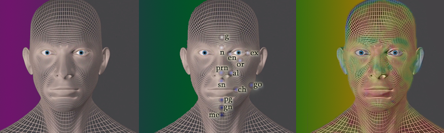
Wize is a mirror that its developers claim can monitor health with breath monitors, 3D scanners, video cameras, and imaging systems. It assesses cardio-metabolic risk through changes in face shape and circulation, signs of anxiety, and  breath tests for heart attack-inducing chemicals.  After a user looks into the mirror for one minute, a health score is produced, and tips for improving health are sent.

Categories
Heart IoT Respiratory Wearables

Phone sensors measure oxygen saturation with out pulse oximeter

MoveSense allows oxygen saturation to be monitored  by phone sensors with what its developers describe as medical accuracy.  A mobile phone must be carried in one’s pocket, and no pulse oximeter is required.  The technology was developed by Bruce Schatz at the University of Illinois.

In a study, patients wore pulse oximeters (for comparison) and carried phones with MoveSense, which continuously recorded saturation and motion. Continuous saturation defined categories corresponding to status levels, including transitions. Continuous motion was used to calculate eight gait parameters from the data. Their existing gait model was then trained with these data points and used to predict transitions in oxygen saturation.

The researchers  discovered that analysis of the saturation, combined with the gait data, could predict saturation with 100 percent accuracy. The model accounts for patients walking faster and slower, which impacts their hearts and lungs.