Categories
Brain Parkinson's

Joe Wang developed, closed-loop, levadopa delivery/monitoring system for Parkinson’s disease

Early Parkinson’s Disease patients benefit significantly from levodopa, to replace dopamine to restore normal motor function. As PD progresses, the brain loses more dopamine-producing cells, which causes motor complications and unpredictable responses to levodopa. Doses must be increased over time, and given at shorter intervals. Regimens are different for each person and may vary from day-to-day.

Currently, clinicians assess levodopa’s benefit by patient testimony and clinical exam, making it difficult to determine optimal treatment. Novel levodopa delivery strategies and wearable sensors that track symptoms and disease progression have been created, but levodopa levels in the body have not been monitored in real time.

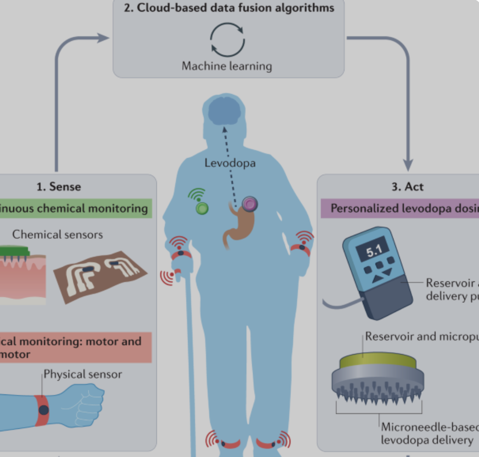
Joe Wang and colleagues have developed a closed loop levodopa delivery system. A network of physical and chemical sensors monitor levodopa levels and inform a delivery device, guided by algorithms, creating a personalized regimen. This can finally optimize the therapeutic management of Parkinson’s Disease.


Join ApplySci at MIT on September 30th for Deep Tech Health + Neurotech Boston
Categories
Brain Sensors

Carbon nanotube sensor precisely measures dopamine

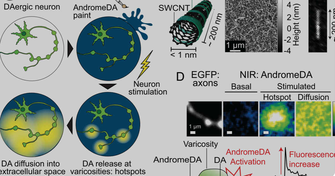
Ruhr University professor Sebastian Kruss, with Max Planck researchers Sofia Elizarova and James Daniel, has developed a sensor that can visualize the release of dopamine from nerve cells with unprecedented resolution. The team used modified carbon nanotubes that glow brighter in the presence of the messenger substance dopamine.

Eizarova said that the sensor “provides new insights into the plasticity and regulation of dopamine signals. In the long term, they could also facilitate progress in the treatment of diseases such as Parkinson’s.”


Join ApplySci at MIT on September 30th for Deep Tech Health + Neurotech Boston
Categories
Brain

Univ. of Reading study links Alzheimer’s disease to blood brain barrier damage

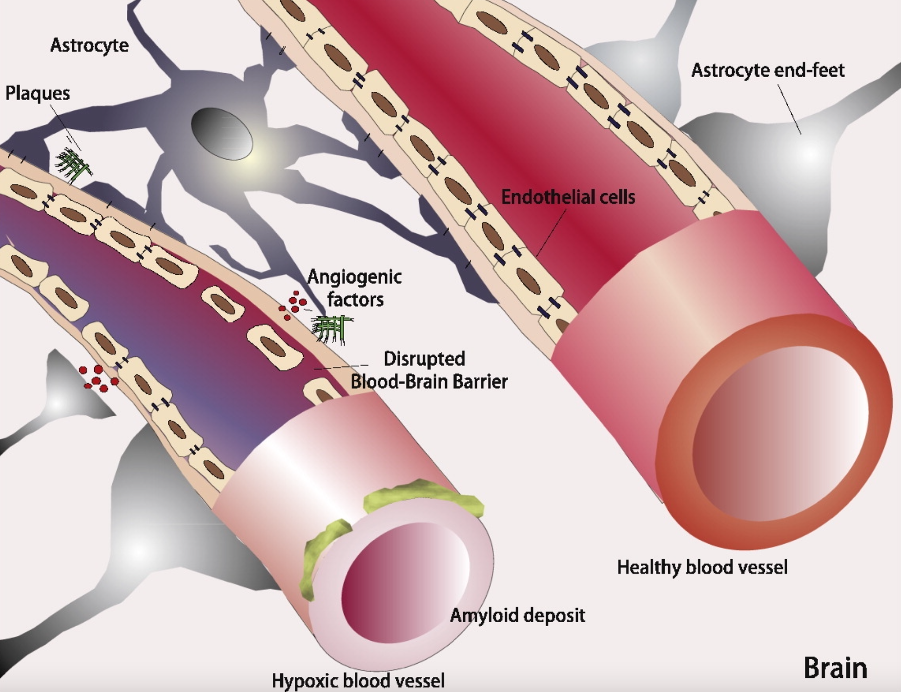
The “Lipid Invasion Model” argues that lipids entering the brain due to blood brain barrier damage is the determining cause of the Alzheimer’s Disease. The presence of excess lipids in the brain cells of Alzheimer’s patients is an element of Alois Alzheimer’s 1906 research, but little has been published about this connection since.

The hypothesis, published in the Journal of Alzheimer’s Disease Reports, could impact diagnosis and treatment, and supports lifestyle changes (ie diet for cholesterol management) to reduce risk.

According to Post Doc Jonathan Rudge, invading lipids can result in brain shrinkage, and amyloid plaque and tau tangle development.

The new study follows 10 years of research and suggests that risk factors associated with Alzheimer’s Disease are the same factors that damage the blood brain barrier—advanced age, head injury, hypertension, smoking, obesity, diabetes, chronic sleep deprivation and stress.


Join ApplySci at MIT on September 30, 2022 for Deep Tech Health + Neurotech Boston
Categories
Autism Brain MRI

Prenatal MRI study suggests autism differences may begin in the womb

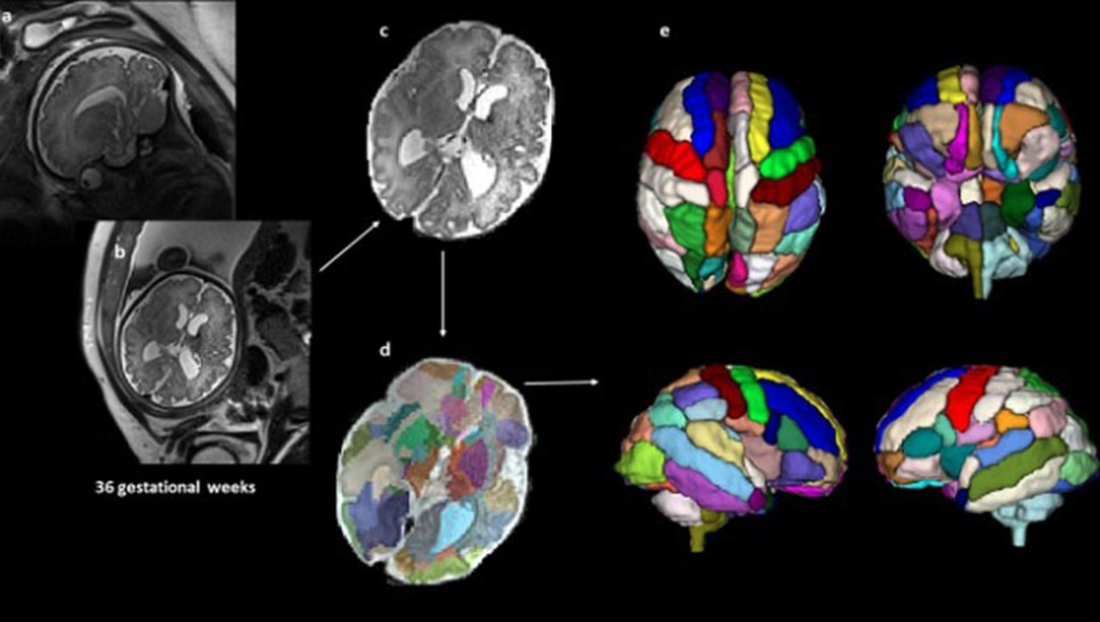
A small Boston Children’s Hospital study led by Assistant Professor Emi Takahashi and postdoc Alpen Ortug showed increased volume of the insular lobe as a potential strong prenatal MRI biomarker that could predict the emergence of ASD later in life. It revealed significant differences in brain structures at 25 weeks’ gestation between children who were later diagnosed with ASD and those who were not.

If validated, this could enable earlier treatment after birth, which could improve outcomes. Early treatment has been shown to improve language and cognitive abilities, but current diagnostic tools can only identify the disorder around 18 months of age.

39 fetal MRI brain scans at 25 weeks’ gestation taken at Boston Children’s Hospital were analyzed. 9 children were later diagnosed with ASD, 20 were neurotypical and 10 did not have ASD but had other conditions also observed in the children with ASD.

An atlas-based automated anatomical labeling method was used to segment the scans and compare brain regions between the different groups. The insular lobe was found to have significantly larger volume in the ASD group compared with the other 3 control groups.

The findings align with studies that have reported changes in the insular cortex in adults with autism, and suggests these differences may begin in the womb. The scans from children with ASD also showed a significantly larger amygdala and hippocampal commissure compared with children who had other health conditions but not ASD.

According to Ortug, “to the best of our knowledge, this is the first attempt to semi-automatically segment the brain regions in the prenatal stage in patients who are diagnosed with autism later and compare different groups of controls.”


Join ApplySci at MIT on September 30th for Deep Tech Health + Neurotech Boston 2022
Categories
Brain

Stanford study: High dose magnetic stimulation eases severe depression

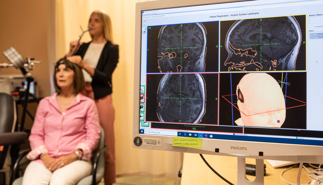
Nolan Williams, Alan Schatzberg, and Stanford colleagues have published a small, double blind study showing that high dose, noninvasive, magnetic brain stimulation alleviated depression symptoms in 80% of participants.

Stanford accelerated intelligent neuromodulation therapy (SAINT) is an intensive, individualized form of transcranial magnetic stimulation. Effects were seen within days and lasted months. Side effects included fatigue and headaches.

29 people with treatment-resistant depression participated. Half received SAINT, and half received a placebo procedure. After five days, 78.6% of the participants in the treatment group had eased symptoms.


See CBS Sunday Morning video describing patient experiences with SAINT


Join ApplySci at MIT on April 8, 2022 for Wearable Tech + Digital Health + Neurotech Boston

Categories
Brain

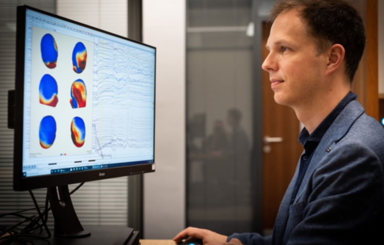
Passive EEG assessment detects cognitive decline early

George Sothart and University of Bath colleagues have developed a new, EEG + game memory assessment technique which could enable the earlier diagnosis of Alzheimer’s disease, the underlying cause of around 60% of dementia cases..

The need for early diagnosis tools to help doctors to prescribe lifestyle interventions to slow the rate of cognitive decline is obvious. This tool could also help identify dementia patients for clinical trials.

“Fastball EEG” is passive. The person performing the test is not given priorinstructions prior to the task, as dementia patients may struggle to follow complex directions, and is not asked to reflect on, respond to or remember any items. She or he simply watches a screen of flashing images.

Two discrete frequency responses are captured, reflecting the participant’s periodic neural responses to the stimuli. The first reflects visual processing; the second mirrors the brain’s response to previously seen images and reflects recognition memory. Analyzing the EEG spectrum at the second, slower frequency can quantify the patient’ memory response.

Fastball EEG was studied in 20 patients with Alzheimer’s disease, 20 healthy older adults and 20 healthy younger adults. For both the recognition and repetition conditions, Fastball EEG detected significantly impaired recognition memory in Alzheimer’s disease patients compared with healthy older control subjects. There were no differences between the two groups under the control condition, where image recognition was not included. The Fastball test could also discriminate Alzheimer’s disease patients from healthy older adult controls, with an accuracy of 86%. No significant performance differences were seen between older and younger healthy controls.

After the Fastball task, participants completed a forced-choice task, in which they had to identify a previously seen image from two alternatives. Here, the researchers observed little difference between Alzheimer’s disease patients and controls, suggesting that Fastball was more sensitive to memory performance than this behavioral recognition test.


Join ApplySci at MIT on April 8, 2022 for Wearable Tech + Digital Health + Neurotech Boston

Categories
Brain

Apple partners with UCLA, Biogen for iPhone mental/cognitive health monitoring


The Apple/UCLA project “Seabreeze” and Apple/Biogen project “Pi” represent a further move into iPhone health monitoring.

According to the Wall Street Journal, Apple is attempting to develop an algorithm to identify depression and cognitive decline from sleep patterns, mobility, and how one uses the phone — for example, how often they look at its clock.

This follows a study that Apple Did with Eli Lilly, which showed that MCI and early dementia patients typed slowly, used devices erratically, sent fewer texts, and used “helper apps” often.

A recent Apple/Duke study analyzed kids phone use to understand emotions and behavior that could be associated with autism.

Mindstrong Health and others have been evaluating phone use to detect mental illness for years, and Mindstrong has recently moved into therapy and psychiatry offerings, based on phone based digital phenotyping, which could be where Apple is headed with these new projects.

The very earliest stages of mental and cognitive wellness changes could be missed using phone data alone, but could still be detected with EEG or other brain signal monitoring technology.

———-

Join ApplySci at MIT on April 8, 2022 for the 14th Wearable Tech + Digital Health + Neurotech Conference.

Categories
BCI

Algorithm isolates specific brain signals, provides feedback

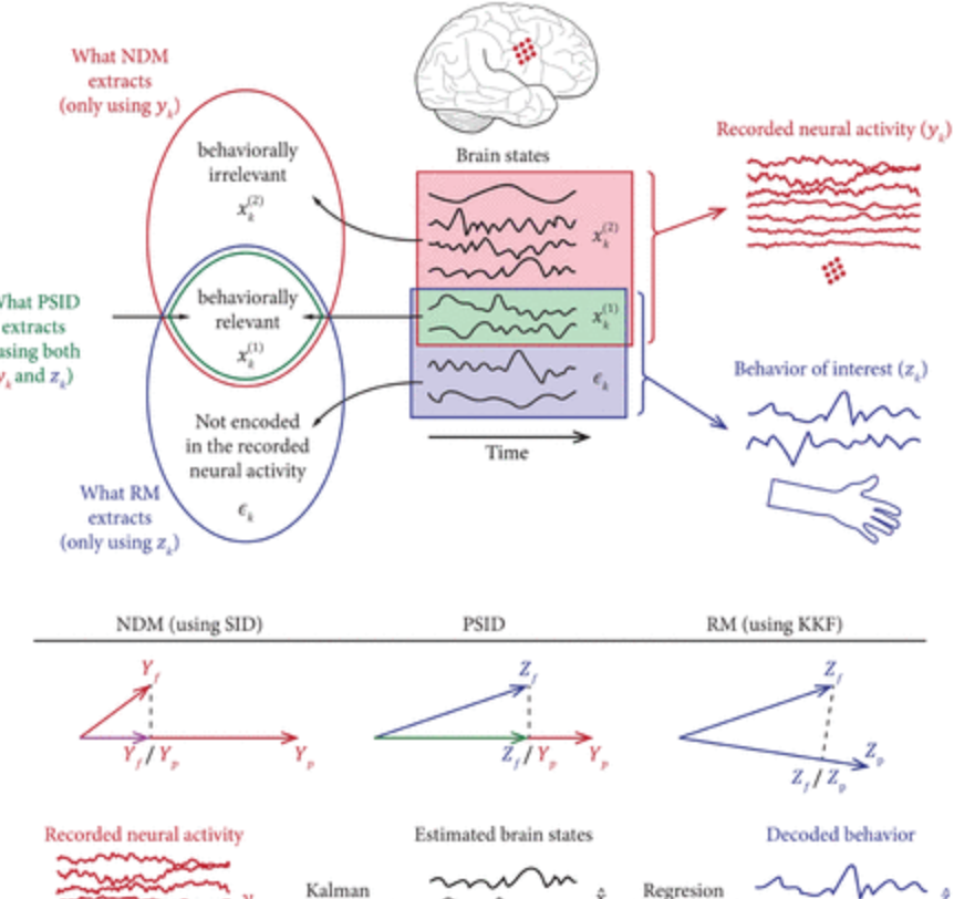
The US Army and USC Prof Maryam Shanechi have developed an algorithm that can determine which specific behaviors—like walking and breathing—belong to specific brain signals.

Segmenting brain signals has been notoriously difficult, as all signals associated with tasks mix together. Shanechi and her team used the algorithm to separate behaviorally relevant brain signals from behaviorally irrelevant brain signals.

Army research office project manager Hamid Krim said that if the algorithm detects behavior indicating a soldier is stressed or overloaded, then a machine could alert that soldier before they recognize their own fatigue. This may enable the development of technology that can interpret signals from the brain and then send signals back, to automatically correct behavior, or to enable soldiers to communicate with out speaking.

In the civilian world, this could allow locked-in patients to communicate, and those with various disabilities and brain diseases to improve function.

Categories
BCI Brain Sensors

Polymer improves medical implants, could enable brain-computer interface

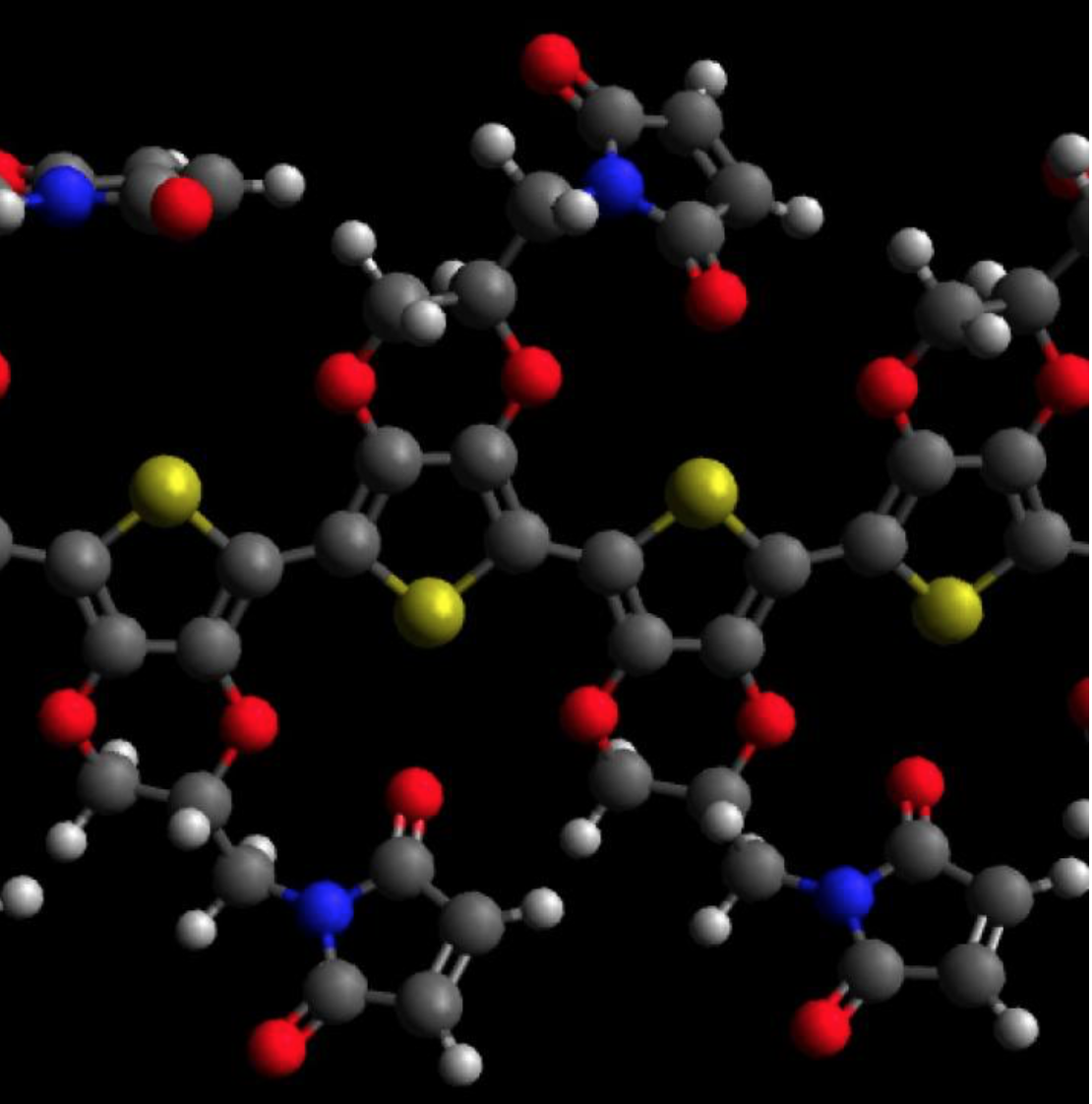
David Martin and University of Delaware colleagues have developed a bio-synthetic coating for electronic components that could avoid the scarring (and signal disruption) caused by traditional microelectric materials. The PEDOT polymer improved the performance of medical implants by reducing their opposition to an electric current.

Pedot film was used with an antibody to stimulate blood vessel growth after injury, and could be used to detect early stages of tumor growth. The polymers could also help sense or treat brain or nervous system disorders, while versions could theoretically attach peptides, antibodies and DNA.

The team believes that materials, when inserted, could connect brains to a computer.

Categories
Brain Sensors

Sensor platform detects dopamine in sweat; could be used for future treatment

Penn State’s Aida Ebrahimi and Maurico Terrones, RPI’s Humberto Terrones, and colleagues, have developed a highly sensitive, non-invasive wearable Dopamine sensor platform. The goal is the use of the technology to develop wearable sensors able to track and eventually treat conditions caused by too much (ie schizophrenia) or too little (ie Parkinson’s, depression) dopamine.

The low cost, flexible detector was achieved by doping a Molybdenum disulfide with Manganes, embedded in two-dimensional transition metal dichalcogenide.

Current dopamine monitoring methods are invasive and require specialized lab equipment. The researchers described the new method as “very simple and scalable.”

Categories
Brain

Brain cholesterol map could lead to neurodegeneration therapies, help guide surgery

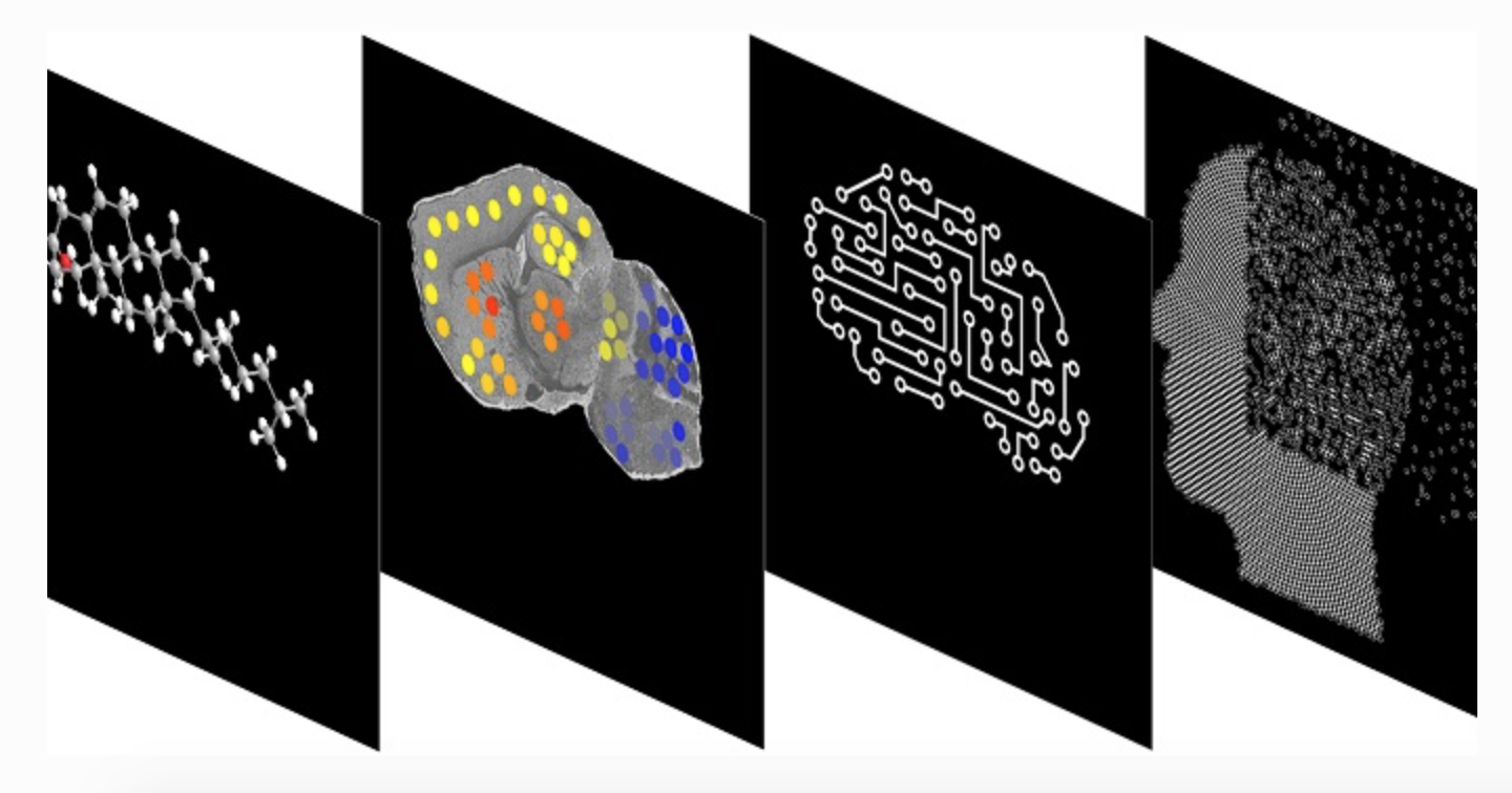
William Griffiths and Swansea University colleagues have developed a method to map cholesterol in the brain, and understand potential molecule conversion. This is the first technology that can map cholesterol metabolism in defined locations at microscopic levels, and visualize how it changes in pathological niches in the brain.

While only studied on mice, the hope is that the technology can also be used to understand brain function changes in humans with neurodegenerative disorders. Griffiths added that “tissue excised during surgery could rapidly be profiled by our method in-clinic and used to distinguish healthy from diseased tissue, informing the surgeon on the next step of the operation.”

Collaborator Yuqin Wang said: “Our results show that cholesterol turnover is particularly high in striatum, the area most affected in Huntington’s disease. We will apply this method to find out how cholesterol metabolism is associated with this disease. This may lead to the development of new therapies to a disease which currently has no cure.”

Categories
Brain EEG

Tattoo electrodes for long-term EEG, MEG measurements

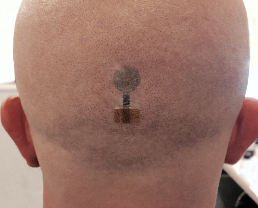
Graz University professor Francesco Greco has built on his earlier work to create advanced inkjet printed conductive polymer electrodes on tattoo paper. The composition and thickness of the transfer paper and conductive polymer have been optimized to achieve a better electrode/skin connection and improve EEG signal quality.

The cheap, user-friendly, dry electrodes have shown similar reliability to traditional EEG electrodes in clinical tests. They can be used for long-term EEG and MEG measurements as they do not contain metal.