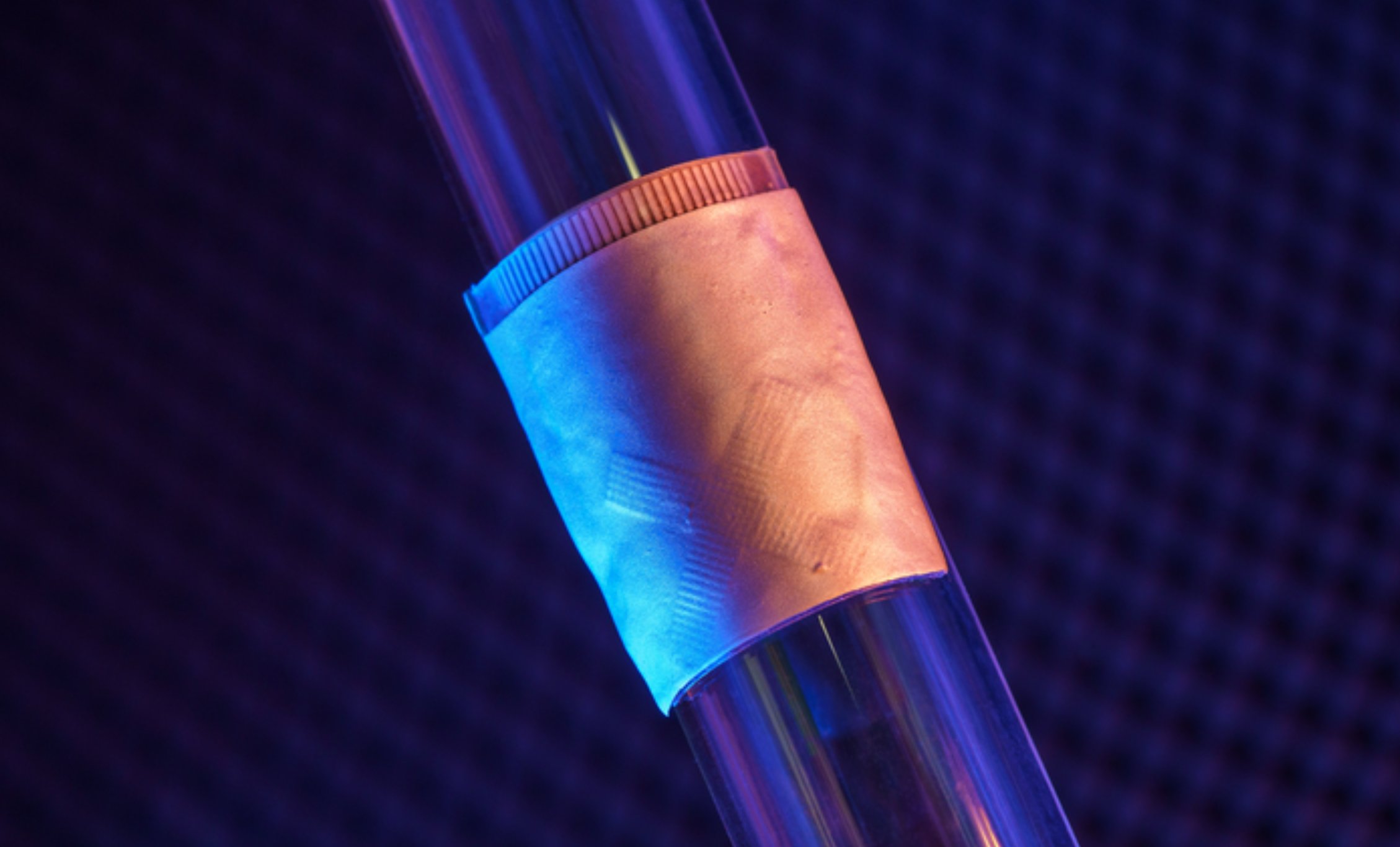
UCSD professor Sheng Xu has developed a wearable ultrasound device that can continuously monitor and assess the structure and function of the human heart for 24 hours, during normal daily activity. This could eliminate the need for highly trained technicians and bulky devices.
Signs of cardiac diseases are transient and unpredictable, and imaging can detect issues before they become problems. The new system gathers information through a small, soft, wearable patch, worn on the chest and designed for optimal adherence. It sends and receives ultrasound waves which are used to generate a constant stream of images of the structure of the heart in real time — with out radiation. As heart function issues often manifest only when the body is in motion, the wearable aspect of the device is particularly important.
Images of the four chambers of the heart are captured in different angles, and a clinically relevant subset of the images is analyzed. The model automatically segments the shape of the left ventricle from the image recording, extracting its volume frame-by-frame and yielding waveforms to measure stroke volume, cardiac output and ejection fraction. Curves of these three indices are generated continuously and noninvasively.
Xu plans to commercialize this technology through Softsonics, a company spun off from UC San Diego.
Sheng Xu was a featured speaker at the 2020 ApplySci Deep Tech Health conference on Sand Hill Road.
