Categories
Cancer Ultrasound

Wearable ultrasound to detect early breast cancer

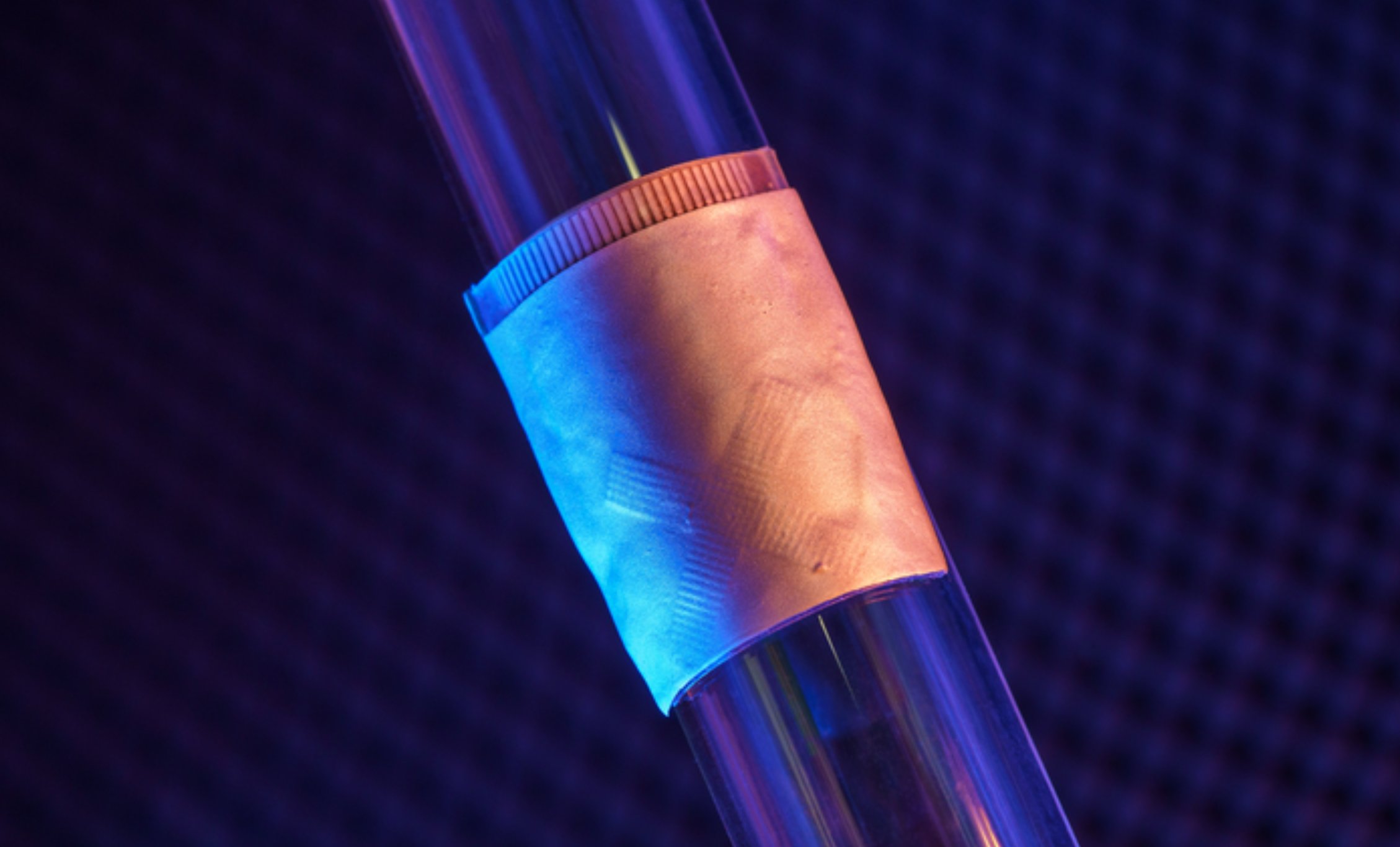
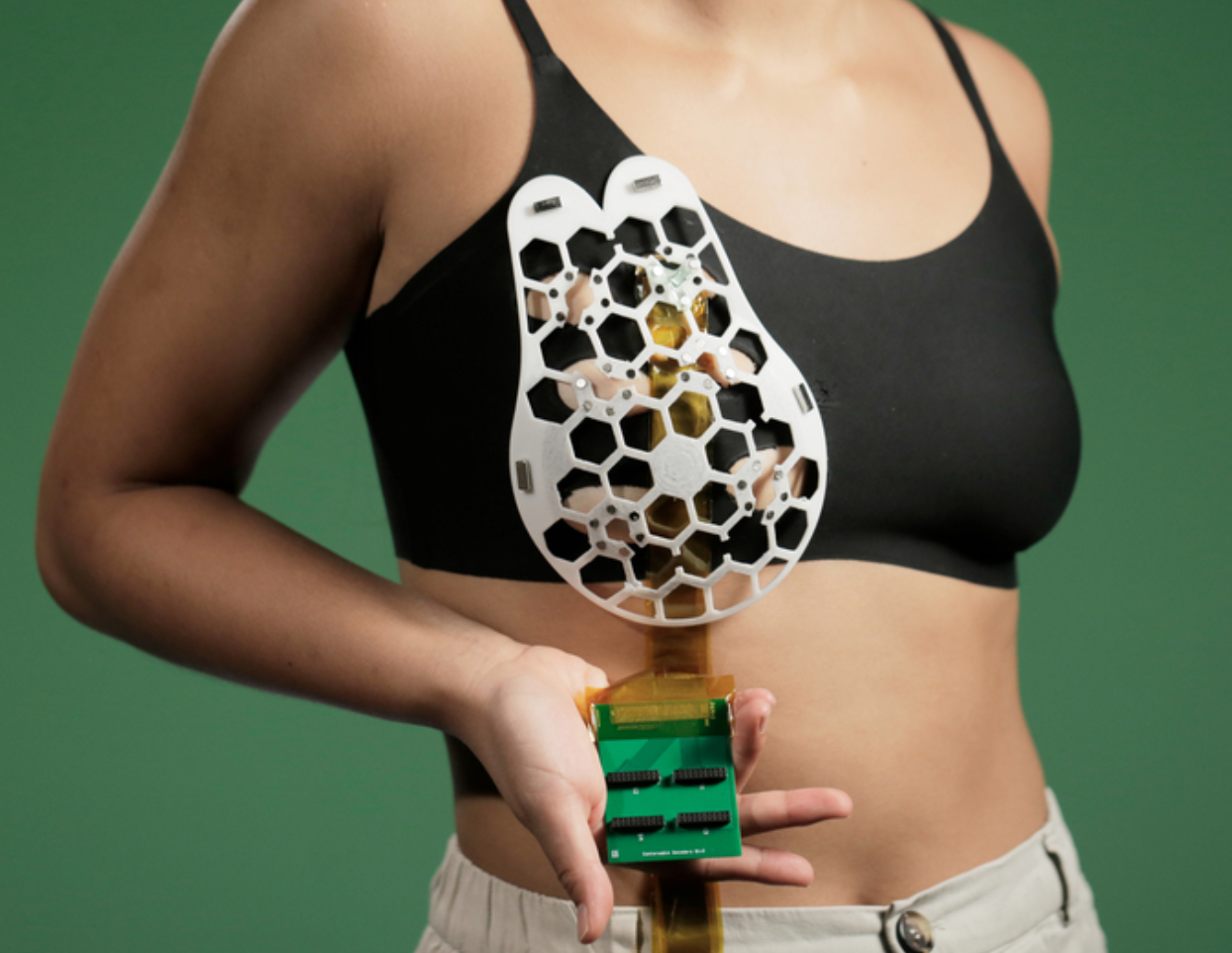
MIT’s Canan Dagdeviren has developed a flexible ultrasound patch that can be attached to a bra, obtaining ultrasound images with resolution comparable to medical imaging centers, and used repeatedly.

Interval cancers, which develop between regularly scheduled mammograms, account for 20 – 30 percent of all breast cancers, and tend to be more aggressive. The goal is to frequently screen those most likely to develop interval cancers.

According to Dagdeviren: “We changed the form factor of the ultrasound technology so that it can be used in your home. It’s portable and easy to use, and provides real-time, user-friendly monitoring of breast tissue.”

Piezoelectric material allowed the scanner to be minimized, in a flexible, 3D-printed patch, with honeycomb shaped openings. Using magnets, it is attached to a bra with openings that allow the it to contact the skin. The scanner fits inside a small tracker, with six different positions, allowing the entire breast to be imaged. IT also rotates, to take images from different angles.

The device was able to detect .3cm diameter cysts in a 71-year-old woman, with a resolution comparable to that of traditional ultrasound. Tissue was imaged at a depth up to 8 centimeters.

To see the images, the scanner must connect to an ultrasound machine, like those used in imaging centers. The team is now working on a miniaturized imaging system.


Join ApplySci at MIT on September 18, 2023 for AI + Deep Tech Health + Neurotech Boston
Categories
Brain Pharmaceuticals Ultrasound

Implanted ultrasound allows powerful chemotherapy drugs to cross the blood brain barrier

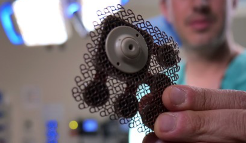
Adam Sonaband and Northwestern colleages used a skull-implantable ultrasound device to open the blood-brain barrier and repeatedly permeate critical regions of the human brain, to deliver intravenous chemotherapy to glioblastoma patients.

This is the first study to successfully quantify the effect of ultrasound-based blood-brain barrier opening on the concentrations of chemotherapy in the human brain. It showed a four- to six-fold increase in concentrations of paclitaxel and carboplatin, powerful drugs not currently used in glioblastoma because they did not cross the blood brain barrier. Temozolomide does cross the blood-brain barrier, but is weaker.

This is also the first study to describe how quickly the blood-brain barrier closes after sonication, allowing optimization of the sequence of drug delivery and ultrasound activation to maximize drug penetration.

The ultrasound implantation takes four minutes, and the patient is awake. It is the basis of an ongoing phase 2 clinical trial.

Categories
Heart Ultrasound Wearables

Wearable cardiac ultrasound continuously monitors heart structure, function for 24 hours

UCSD professor Sheng Xu has developed a wearable ultrasound device that can continuously monitor and assess the structure and function of the human heart for 24 hours, during normal daily activity. This could eliminate the need for highly trained technicians and bulky devices. 

Signs of cardiac diseases are transient and unpredictable, and imaging can detect issues before they become problems. The new system gathers information through a small, soft, wearable patch, worn on the chest and designed for optimal adherence. It sends and receives ultrasound waves which are used to generate a constant stream of images of the structure of the heart in real time — with out radiation. As heart function issues often manifest only when the body is in motion, the wearable aspect of the device is particularly important.

Images of the four chambers of the heart are captured in different angles, and a clinically relevant subset of the images is analyzed. The model automatically segments the shape of the left ventricle from the image recording, extracting its volume frame-by-frame and yielding waveforms to measure stroke volume, cardiac output and ejection fraction. Curves of these three indices are generated continuously and noninvasively.

Xu plans to commercialize this technology through Softsonics, a company spun off from UC San Diego.


Sheng Xu was a featured speaker at the 2020 ApplySci Deep Tech Health conference on Sand Hill Road.

Categories
Brain Conference Parkinson's Ultrasound

Focused ultrasound thalamotomy in Parkinson’s Disease

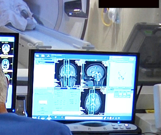
UVA’s Scott Sperling and Jeff Elias, who already used focused ultrasound to treat essential tremor, have just published the results of  a small study showing the efficacy of the technology in Parkinson’s Disease.

The sound waves were shown to interrupt brain circuits responsible for the uncontrollable shaking associated with the disease. The researchers claim that their study also offers “comprehensive evidence of safety” in its effect on mood, behavior and cognitive ability, which has not previously been studied.

According to Sperling, “In this study, we extended these initial results and showed that focused ultrasound thalamotomy is not only safe from a cognitive and mood perspective, but that patients who underwent surgery realized significant and sustained benefits in terms of functional disability and overall quality of life.”

27 adults with severe Parkinson’s tremor that had not responded to previous treatment were divided  into two groups. Twenty received the procedure, and a control group of seven (who were later offered the procedure) did not. Participants reported improved quality of life, including their ability to perform simple daily tasks, emotional wellbeing, and a lessened sense of stigma due to their tremor, at both three and twelve months.

The team found that mood and cognition, and the ability to go about daily life, ultimately had more effect on participants’ assessment of their overall quality of life than did remor severity or the amount of tremor improvement.

Cognitive decline was seen in some participants after the study, in that they were less able to name colors and think of and speak words. The cause of this was unclear, and must be investigated. The researchers suggested this could be a result of the natural progression of Parkinson’s.


Join ApplySci at the 10th Wearable Tech + Digital Health + Neurotech Silicon Valley conference on February 21-22 at Stanford University — Featuring:  Zhenan BaoChristof KochVinod KhoslaWalter Greenleaf – Nathan IntratorJohn MattisonDavid EaglemanUnity Stoakes Shahin Farshchi Emmanuel Mignot Michael Snyder Joe Wang – Josh Duyan – Aviad Hai Anne Andrews Tan Le