Categories
AI

Google AI detects tuberculosis

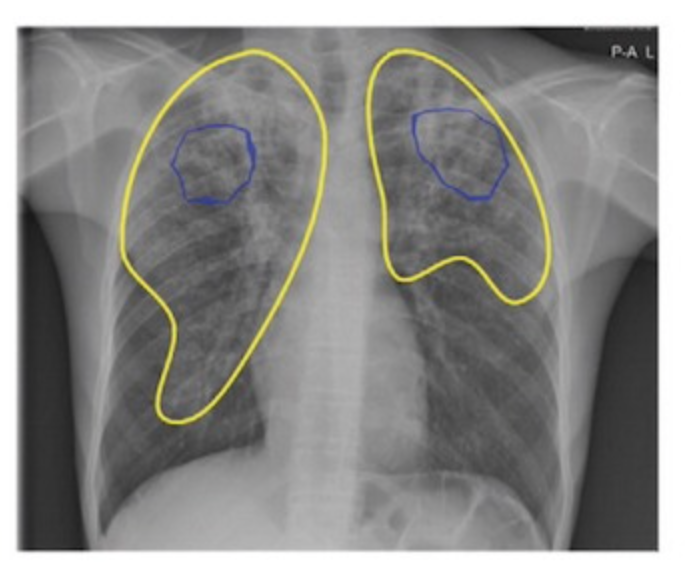
Google’s deep learning technology detected tuberculosis with similar accuracy to radiologists in a Radiology study.

165,174 chest radiographs from 22,284 patients in four countries were scanned. In detecting active tuberculosis, its sensitivity was higher (88 percent versus 75 percent) and its specificity was noninferior (79 percent versus 84 percent) compared to nine radiologists. Costs were reduced by 40 to 80 percent per tuberculosis-positive patient.

This may be able to facilitate tuberculosis screening in areas with limited radiologist resources, improving public health.


Join ApplySci at MIT for Deep Tech Health + Neurotech Boston on September 30th, with talks by:

Giovanni Traverso – Bob Langer – Emery Brown – George Church – Mary Lou Jepsen – Tom Oxley – Ellen Roche – Nathan Intrator – Bakul Patel – Ramita Tandon – Shaun Patel – Elizabeth Andukowich – Cris De Luca – Robert Garber – Jonathan Behr – Ann DeWitt

Categories
Brain Parkinson's

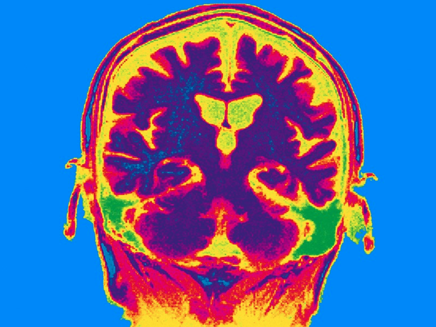
qMRI for early detection of Parkinson’s disease

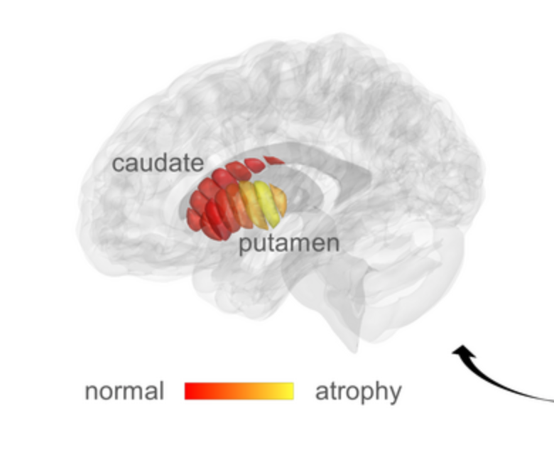
Aviv Mezer and Hebrew University colleagues used quantitative MRI to identify cellular changes in Parkinson’s disease. Their method enabled them to look at microstructures in the striatum, which is known to deteriorate during disease progression. Using a novel algorithm developed by Elior Drori, biological changes in the striatum were revealed, and associated with early stage Parkinson’s, and movement dysfunction.

qMRI achieves its sensitivity by taking several MRI images using different excitation energies. The researchers used this to reveal changes in the tissue structure within distinct regions of the striatum. Previously, the structural sensitivity of these measurements could only be seen post mortem. 

Mezer’s goal is to facilitate early diagnosis of the disease, and provide markers for monitoring the efficacy of future therapies. He also seeks to identify subgroups within the population suffering from Parkinson’s disease – who may respond differently to some drugs than others, leading to personalized treatment. He will now use the technique to investigate microstructural changes in other regions of the brain. The team is in the early stages of developing a qMRI into a tool that can be used in a clinical setting.


Join ApplySci at MIT on September 30 for Deep Tech Health + Neurotech Boston

Categories
Brain

Non-invasive stimulation improves memory in study

In a recent study, Boston University professor Robert Reinhart used tACS to stimulate brain activity in 150 people aged 65-88, resulting in memory improvements for one month.

Stimulating the dorsolateral prefrontal cortex improved long-term memory, while stimulating the inferior parietal lobe, with low-frequency electrical currents, boosted working memory.

Participants were asked to recall 20 words that were read aloud. They underwent tACS for the entire 20 minute duration of the task.After four consecutive days, participants who received high-frequency stimulation of the dorsolateral prefrontal cortex had an improved ability to remember words from the beginning of the lists, which depends on long-term memory. Low-frequency stimulation of the inferior parietal lobe enhanced participants’ recall of items later in the lists, which involves working memory. Performance improved over the four days — and the gains persisted a month later. Those who had the lowest levels of general cognitive function before the study experienced the largest memory improvements.

Changing frequencies and brain regions (applying high-frequency stimulation to the parietal lobe, for instance), or using a ‘sham’ protocol in which the electrical currents were applied only briefly at the beginning and end of the task to mimic the sensation of brain stimulation, did not boost memory.

The team is exploring the use of tACS in Alzheimer’s disease, as the study indicated that brain stimulation might provide the greatest benefits to those who have poor cognitive function.


Join ApplySci at MIT for Deep Tech Health + Neurotech Boston on September 30th, with talks by:

Giovanni Traverso – Bob Langer – Emery Brown – George Church – Mary Lou Jepsen – Tom Oxley – Ellen Roche – Nathan Intrator – Bakul Patel – Ramita Tandon – Shaun Patel – Elizabeth Andukowich – Cris De Luca – Robert Garber – Jonathan Behr – Ann DeWitt

Categories
AI Brain Parkinson's

Neural Network assesses sleep patterns for passive Parkinson’s diagnosis

MIT’s Dina Katabi has developed a non-contact, neural network-based system to detect Parkinson’s disease while a person is sleeping.

By assessing nocturnal breathing patterns, the series of algorithms detects, and tracks the progression of, the disease — every night, at home.

A device in the bedroom emits radio signals, analyzes their reflections off the surrounding environment, and extracts breathing patterns, without bodily contact. The breathing signal is then fed to the neural network to assess Parkinson’s Disease in a passive manner.

Current diagnosis methods are invasive, expensive, and must be done at specialized centers, making frequent testing almost impossible.

Katabi said that a relationship between Parkinson’s and breathing was noted in 1817, motivating her to explore this form of detection, and that respiratory symptoms manifest years before motor symptoms.


Join ApplySci at MIT on September 30th for Deep Tech Health + Neurotech Boston

Categories
AI Cancer

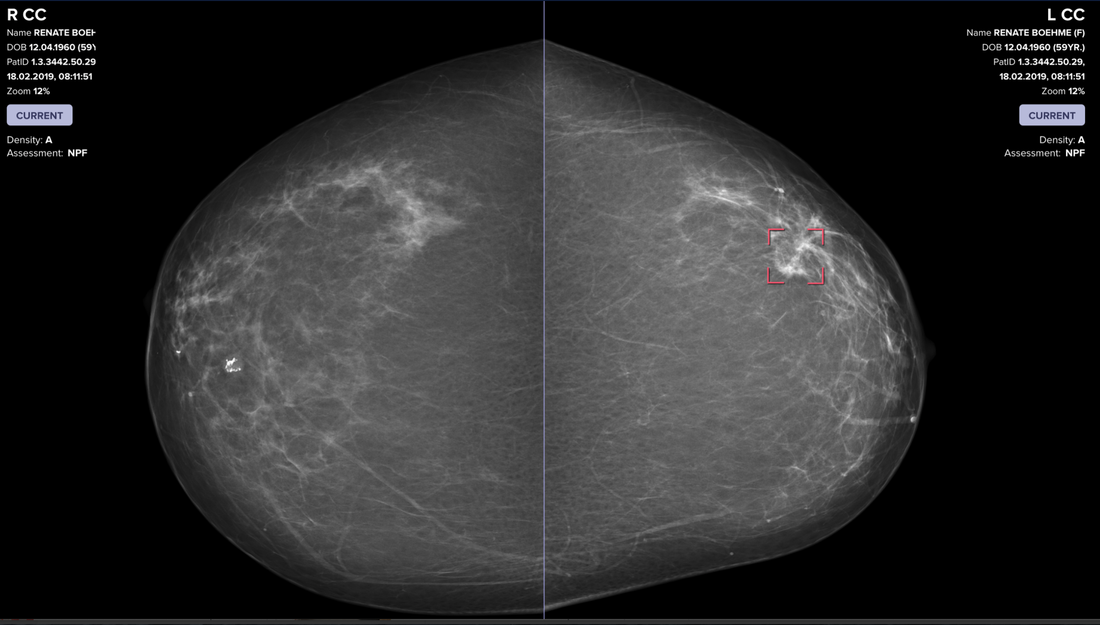
AI catches breast cancer earlier, more often than traditional screening alone

The mammography screening paradigm has not changed since the 1960s.

Breast screening AI company Vara, with Essen University and Memorial Sloan Kettering hospitals, published a study showing that radiologists assisted by AI are better able to screen for breast cancer. The hope is that AI systems could detect cancers that doctors miss, provide better care in remote areas, and allow radiologists more time to see more patients.

Two approaches were tested. In the first, AI analyzed mammograms. In the second, AI distinguished between normal and concerning scans. It refers the latter to a radiologist, who reviews them before seeing the AI’s assessment. Then the AI issues a warning if it detected cancer when the doctor did not. In the study, the AI examined old scans and compared its assessments with those of the radiologist who reviewed them.

Data from 367,000 mammograms—including radiologists’ notes, original assessments, and information on whether the patient ultimately had cancer— was analyzed to learn how to place these scans into one of three categories: “confident normal,” “not confident” (in which no prediction is given), and “confident cancer.” The conclusions from both approaches were then compared with the decisions real radiologists originally made on 82,851 mammograms sourced from screening centers that didn’t contribute scans used to train the AI.

The second approach—doctor and AI working together—was 2.6% better at detecting breast cancer than a doctor working alone, and raised fewer false alarms. It automatically set aside scans it classified as “confident normal,” which was 63% of all mammograms.


Clairity, an AI-driven precision breast cancer screening company, using Harvard professor Connie Lehman and colleagues’ decades of research in radiomics and applied AI, has developed a rigorous scientific approach to improve the accuracy of risk assessment. Prof Lehman discussed her approach at the 2019 ApplySci conference at Harvard Medical School.


Join ApplySci at MIT on September 30, 2022 for Deep Tech Health + Neurotech Boston featring Giovanni Traverso, MITBakul Patel, GoogleGeorge Church, Harvard Kerri Dugan, DARPAEllen Roche, MITNathan Intrator, NeurosteerEmery Brown, HarvardMary Lou Jepsen, OpenwaterShaun Patel, REACT NeuroElizabeth Andukowich, NIMHRamita Tandon, WalgreensCris de Luca, Sanofi VenturesTom Oxley, Synchron

Categories
Respiratory Sensors Wearables

Small sticker-sensor continuously analyzes breath for broad health monitoring

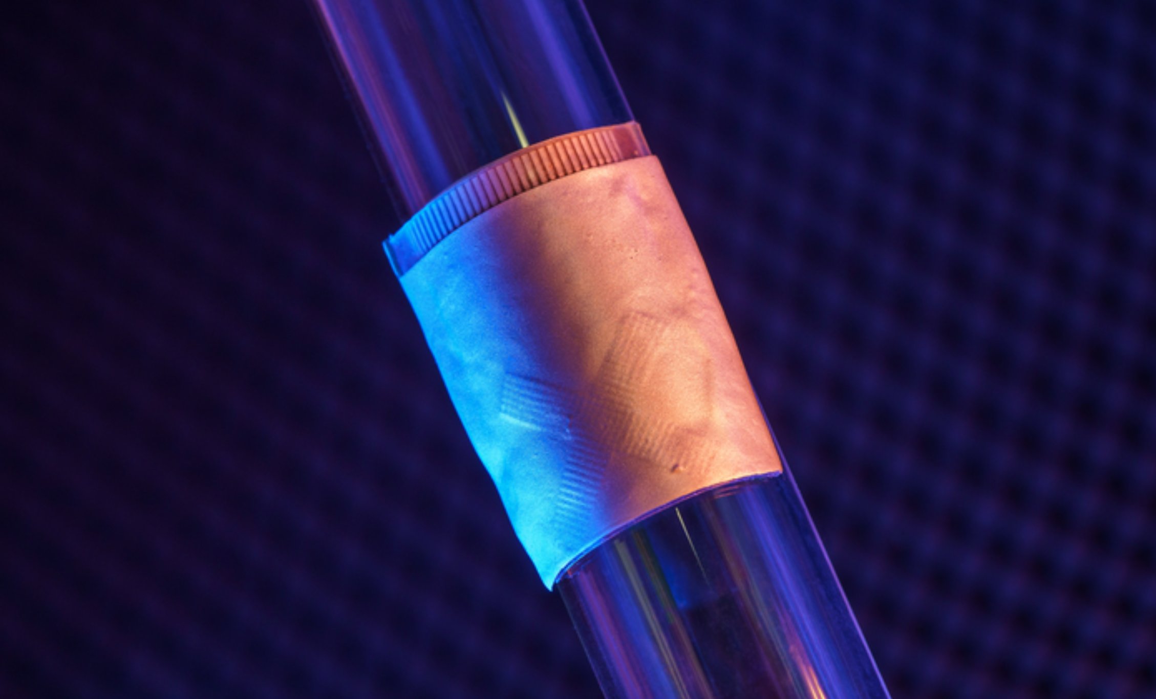
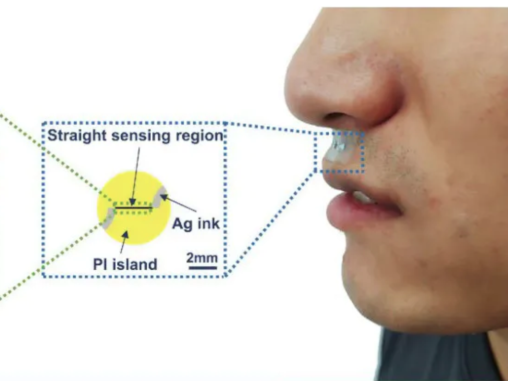
Heibei University, Tianjin Hospital, Beihang University, and Penn State researchers have developed an under nose-worn, stretchable, skin-friendly, waterproof sensor to analyze breath for health monitoring. It could be use for multiple-condition screening, asthma and COPD management, or environmental hazard sensing, among other applications.

The functional gas sensor, in a moisture-resistant membrane, can operate in humid environments, such as below the nose, to monitor breath. It is skin-friendly, stretching, twisting, and conforming to the skin.


Join ApplySci at MIT on September 30th for Deep Tech Health + Neurotech Boston

Categories
Pregnancy Sensors

Remote, non-invasive pregnancy monitor tracks uterine activity

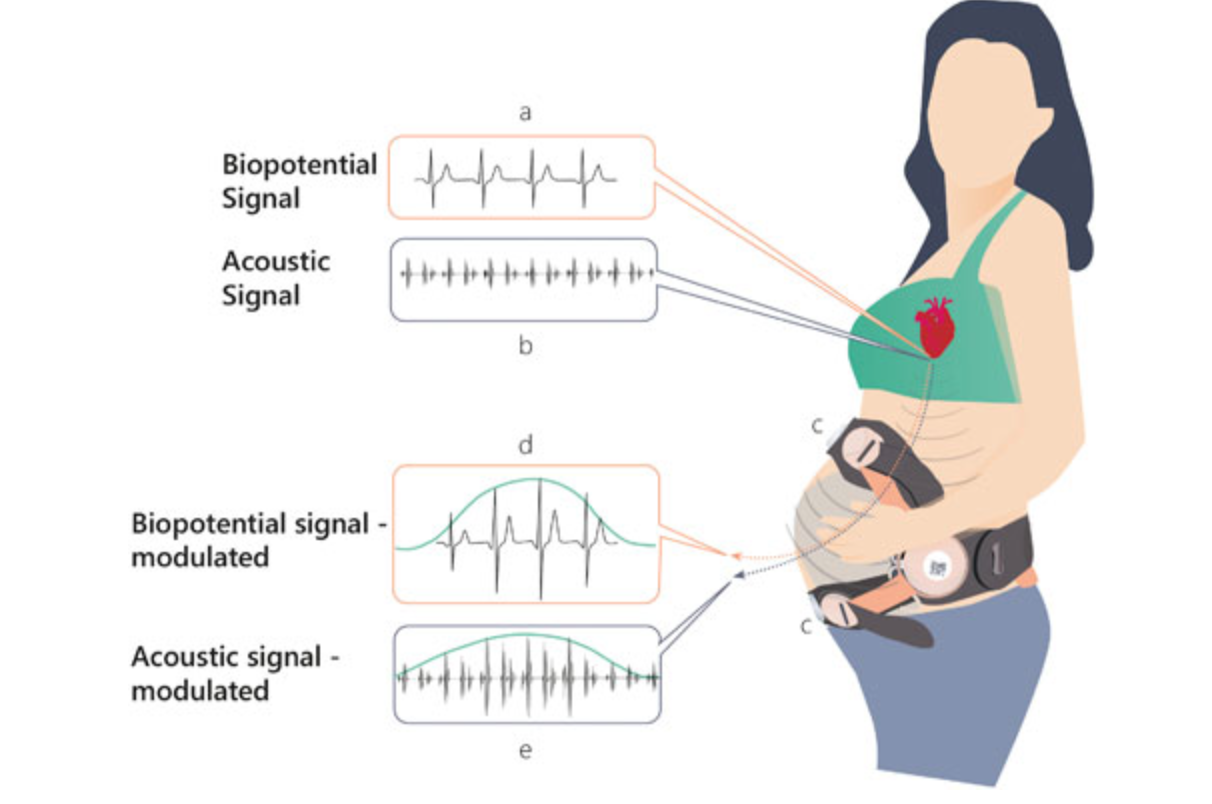
Uterine activity monitoring is essential to pregnancy management. Current methods are either invasive, or their accuracy is compromised by obesity, maternal movements, or belt positioning.

Pregnancy-monitoring company Nuvo has published a study showing that their cardiac-derived algorithm for uterine monitoring was more accurate than TOCO standard of care in 150 patients. This remote, non-invasive detection and monitoring of UA builds on the company’s existing remote fetal heart rate monitoring capabilities. Their goal is to bring comprehensive pregnancy monitoring home.


Join ApplySci at MIT on September 30th for Deep Tech Health + Neurotech Boston

Categories
BCI Brain

First US patient receives Synchron endovascular BCI implant

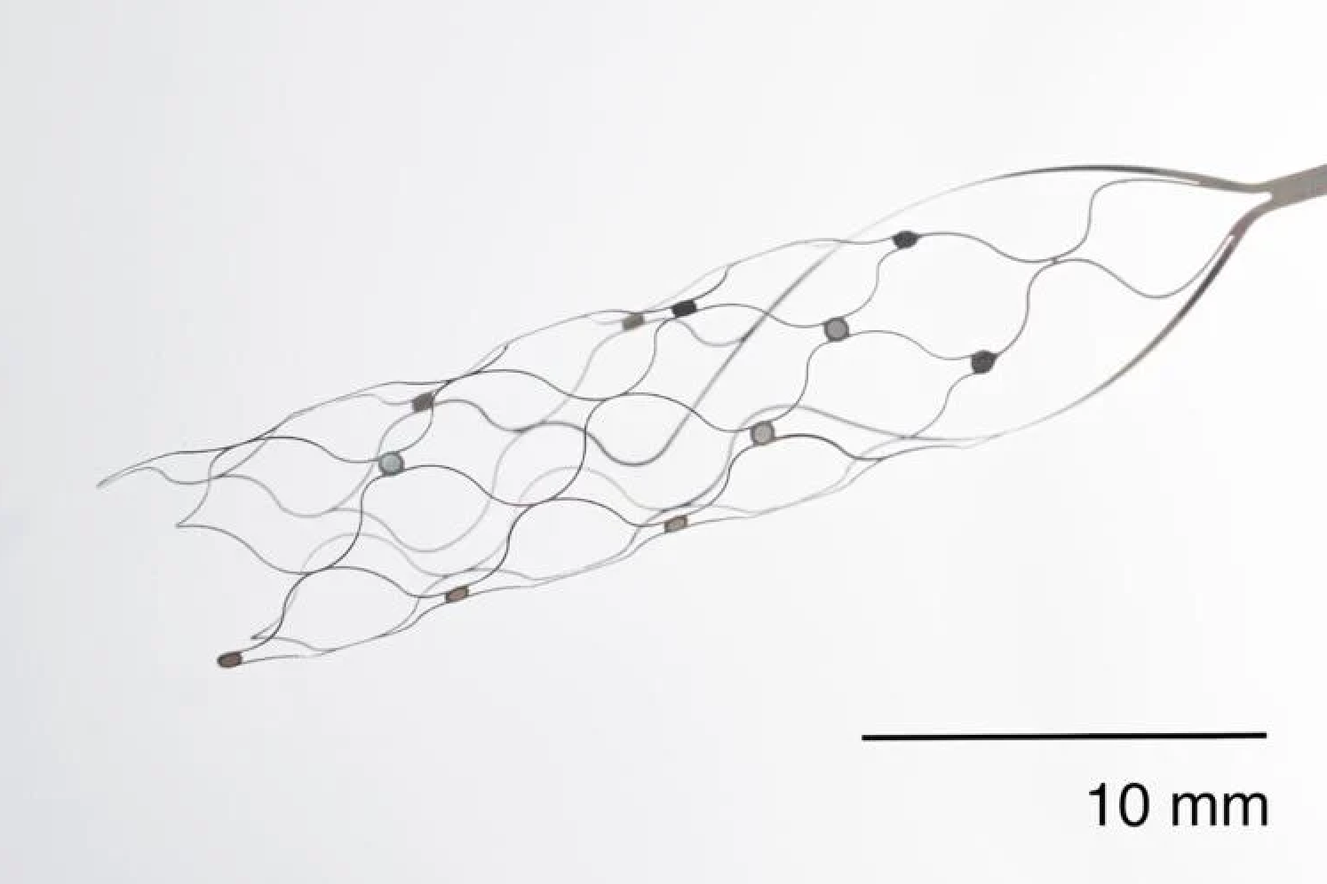
On July 6, 2022, Mount Sinai’s Shahram Majidi threaded Synchron‘s 1.5-inch-long, wire and electrode implant into a blood vessel in the brain of a patient with ALS. The goal is for the patient, who cannot speak or move, to be able to surf the web and communicate via email and text, with his thoughts.

Four patients in Australia have already received the Synchron implant. They have not had side effects, and have been able to send WhatsApp messages and make online purchases.

The “Stentrode” device can be inserted into the brain without cutting through a person’s skull or damaging tissue. An incision is made in the neck, and the stentrode is fed, via catheter, through the jugular vein, into a blood vessel within the motor cortex. As the catheter is removed, the stentrode opens and begins to fuse with the outer edges of the vessel. The procedure takes a few minutes.

A second procedure connects the stentrode to a computing device in the patient’s chest, with a wire. A surgeon creates a tunnel for the wire and a pocket for the device underneath the patient’s skin, similar to a pacemaker procedure. The stentrode reads neuron signals, and the computing device amplifies them and sends them to a computer or phone via Bluetooth.

Synchron aims to shrink the size of its devices, and increase their computing power. It hopes to be able to place numerous stentrodes in different parts of the brain, allowing the patient toperform more functions.

The company was founded by Dr. Thomas Oxley, who will be a featured speaker at ApplySci’s Deep Tech Health + Neurotech conference at MIT on September 30, 2022.

Categories
Babies Brain Sensors

Sensor jumpsuit monitors infant motor abilities


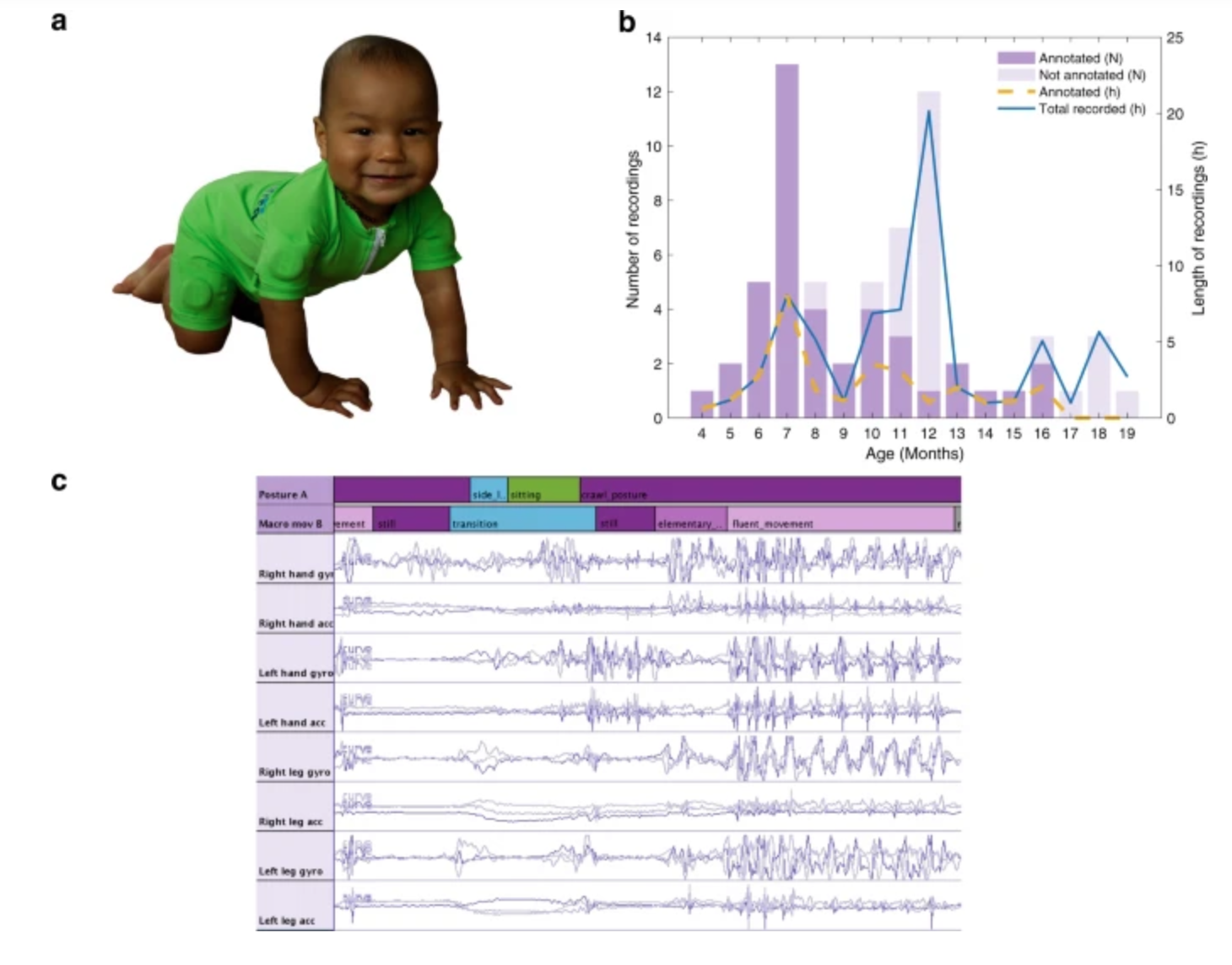
Sampsa Vanhatalo, Manu Airaksinen and University of Helsinki colleagues have developed MAIJU (Motor Assessment of Infants with a Jumpsuit,) a wearable onesie with multiple movement sensors which they believe is able to predict a child’s neurological development.

In a recent study, 5 to 19 month-old infants were monitored using MAIJU during spontaneous playtime. Initially, infant postures and movements were identified visually from a video using a motility description scheme. This was used to train an algorithm to recognize the same postures and movements for every second of each child’s playtime, making it possible to assess her or him in a natural environment.

The goal is the earliest possible detection of neurodevelopmental delays, for earlier intervention. and better outcomes, as therapies would be a part of the child’s everyday life and environment.

The researcher believe that their technology could be automized and effectively adapted to help older children and seniors.

Click to view University of Helsinki video


JOIN APPLYSCI at MIT on SEPTEMBER 30th for Deep Tech Health + Neurotech

Giovanni Traverso – MIT |  George Church – Harvard, MIT | Kerri Dugan – DARPA |  Emery Brown – Harvard |  Bakul Patel – Google |  Nathan Intrator – Neurosteer|  Ramita Tandon – Walgreens |  Ellen Roche – MIT |  Shaun Patel – REACT Neuro | Tom Oxley – Synchron |  Elizabeth Ankudowich – NIH |  Cris De Luca – Sanofi Ventures |  Mary Lou Jepsen – Openwater |  Bob Langer – MIT

Categories
Blood Pressure Sensors

Continuous, cuffless blood pressure monitoring via graphene tattoo

Deji Akinwande, Roozbeh Jafari, and UT Austin colleagues have developed an electronic wrist tattoo that can be worn for hours and deliver highly accurate, continuous blood pressure measurements.

This can provide a much clearer picture of a person’s health than occasional, cuff based measurements at a physicians office, or at home.

Smart watches are not able to successfully measure blood pressure, as they move, are far from arteries, and light-based measurements are often not accurate in people with dark skin or large wrists.

As Akiwande said, “blood pressure is the most important vital sign you can measure” and as Jafari said, “taking infrequent blood pressure measurements has many limitations, and it does not provide insight into exactly how our body is functioning.”

A constant, passive measure, through a tattoo, during normal activity, stress, sleep, or exercise can deliver thousands of measurements more than any existing device.


JOIN APPLYSCI at MIT on SEPTEMBER 30th for Deep Tech Health + Neurotech

Giovanni TraversoMIT | Kerri DuganDARPA | Emery BrownHarvard | Bakul Patel Google | Nathan IntratorNeurosteer | Ramita TandonWalgreens | Ellen RocheMIT | Shaun PatelREACT Neuro | Connie LehmanHarvard | Tom OxleySynchron | Elizabeth AnkudowichNIH | Cris De LucaSanofi Ventures | Mary Lou JepsenOpenwater | Bob LangerMIT

Categories
Brain Parkinson's

Joe Wang developed, closed-loop, levadopa delivery/monitoring system for Parkinson’s disease

Early Parkinson’s Disease patients benefit significantly from levodopa, to replace dopamine to restore normal motor function. As PD progresses, the brain loses more dopamine-producing cells, which causes motor complications and unpredictable responses to levodopa. Doses must be increased over time, and given at shorter intervals. Regimens are different for each person and may vary from day-to-day.

Currently, clinicians assess levodopa’s benefit by patient testimony and clinical exam, making it difficult to determine optimal treatment. Novel levodopa delivery strategies and wearable sensors that track symptoms and disease progression have been created, but levodopa levels in the body have not been monitored in real time.

Joe Wang and colleagues have developed a closed loop levodopa delivery system. A network of physical and chemical sensors monitor levodopa levels and inform a delivery device, guided by algorithms, creating a personalized regimen. This can finally optimize the therapeutic management of Parkinson’s Disease.


Join ApplySci at MIT on September 30th for Deep Tech Health + Neurotech Boston

Categories
Brain Sensors

Carbon nanotube sensor precisely measures dopamine

Ruhr University professor Sebastian Kruss, with Max Planck researchers Sofia Elizarova and James Daniel, has developed a sensor that can visualize the release of dopamine from nerve cells with unprecedented resolution. The team used modified carbon nanotubes that glow brighter in the presence of the messenger substance dopamine.

Eizarova said that the sensor “provides new insights into the plasticity and regulation of dopamine signals. In the long term, they could also facilitate progress in the treatment of diseases such as Parkinson’s.”


Join ApplySci at MIT on September 30th for Deep Tech Health + Neurotech Boston