Categories
Brain

Fluorescent tags allow researchers to observe molecules becoming memories

https://www.einstein.yu.edu/news/releases/968/watching-molecules-morph-into-memories/

Albert Einstein College of Medicine researchers have used advanced imaging techniques to observe memory making molecules travel in real time in living brain cells.

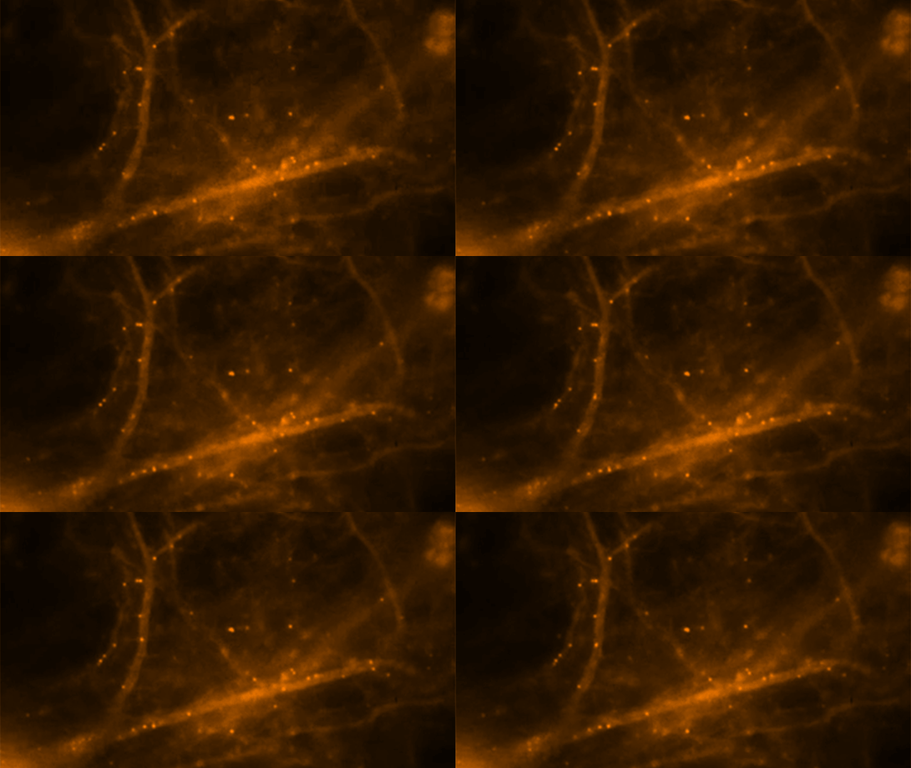
To look deeply into neurons without harming them, the researchers developed a mouse model in which they fluorescently tagged all molecules of messenger RNA that code for beta-actin protein – an essential structural protein found in large amounts in brain neurons and considered a key player in making memories. mRNA is a family of RNA molecules that copy DNA’s genetic information and translate it into the proteins that make life possible. They stimulated neurons from the mouse’s hippocampus, where memories are made and stored, and then watched fluorescently glowing beta-actin mRNA molecules form in the nuclei of neurons and travel within dendrites, the neuron’s branched projections. They discovered that mRNA in neurons is regulated through a novel process described as “masking” and “unmasking,” which allows beta-actin protein to be synthesized at specific times and places and in specific amounts.

Neurons come together at synapses, where slender dendritic “spines” of neurons grasp each other, much as the fingers of one hand bind those of the other. Evidence indicates that repeated neural stimulation increases the strength of synaptic connections by changing the shape of these interlocking dendrite “fingers.” Beta-actin protein appears to strengthen these synaptic connections by altering the shape of dendritic spines. Memories are thought to be encoded when stable, long-lasting synaptic connections form between neurons in contact with each other.

Hye Yoon Park stimulated individual hippocampal neurons of the mouse and observed newly formed beta-actin mRNA molecules within 10 to 15 minutes, indicating that nerve stimulation had caused rapid transcription of the beta-actin gene. Further observations suggested that these beta-actin mRNA molecules continuously assemble and disassemble into large and small particles, respectively. These mRNA particles were seen traveling to their destinations in dendrites where beta-actin protein would be synthesized.

Adina Buxbaum showed that neurons may be unique among cells in how they control the synthesis of beta-actin protein.  This revealed the mechanism by which brain neurons handle this challenge. She found that as soon as beta-actin mRNA molecules form in the nucleus of hippocampal neurons and travel out to the cytoplasm, the mRNAs are packaged into granules and so become inaccessible for making protein. She then saw that stimulating the neuron caused these granules to fall apart, so that mRNA molecules became unmasked and available for synthesizing beta-actin protein.
Categories
BCI Brain

Neurofeedback alters brain plasticity in PTSD

1. Tel Aviv University paper   2. Schulich School of Medicine paper

Professor Ruth Lanius and colleagues at Western University’s Schulich School of Medicine have published a study showing that neurofeedback can improve subjective wellbeing in PTSD, potentially leading to new treatment options. Schulich researchers claim that this is “the first study to show that key brain networks involved in mediating affect and cognition in PTSD can be volitionally modulated via neurofeedback, with measurable outcomes on subjective well-being. It was achieved by harnessing multiple imaging techniques, including EEG and fMRI. Using fMRI we captured the patients’ resting-state brain activity just before and after a 30-minute neurofeedback training session, which was carried out outside the scanner using EEG. We then searched for any differences in connectivity within well-known brain networks. Interestingly, we discovered significant correlations between EEG and fMRI network activities as well as changes in self-reported calmness. This indicated that neurofeedback was able to directly modulate the brain bases of emotional processing in PTSD.”

A group at the Functional Brain Center at Tel Aviv University is trying to achieve similar results using EEG neurofeedback only.  Yehudit Meir-Hasson, Professor Talma HendlerProfessor Nathan Intrator and colleagues published a recent paper demonstrating the validation of an emotional EEG fingerprint with fMRI in the journal NeuroImage.

Categories
AI BCI Brain Robotics

Collaborative cloud system for human-serving robots

http://www.roboearth.org/what-is-roboearth

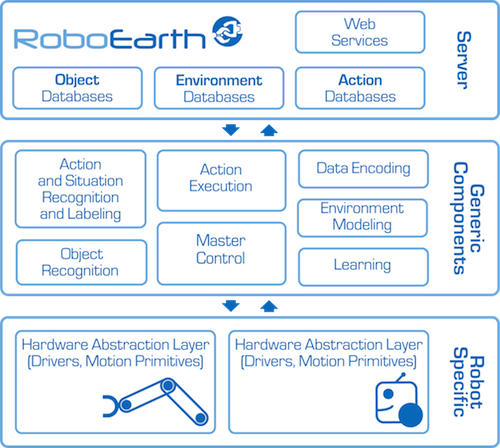
RoboEarth’s goal is to speed the development of human-serving robots. Scientists from five European universities gathered this week for its launch and demonstrated potential applications. This included a robot scanning a room’s physical layout, including the location of the patient’s bed, and the placing a carton of milk on a nearby table.

The system is sometimes called a “Wikipedia” for robots, allowing them, or their programmers,  to share and retrieve information.

Researchers claim that the robots’ technically sophisticated capabilities are comparable to those of high-end robots in car factories. They, however,  look clumsier because robots that interact with humans are not performing repetitive tasks in the controlled, sanitized and predictable surroundings of a factory.

RoboEarth’s system of networked computers enable it to perform intensive computing tasks that smaller computers (or simpler robots) cannot.  It also allows individual robots to communicate between themselves through its “RoboCloud” and robot database. This could facilitate light and cheap robots that use the cloud for computing.

Categories
Brain Eyes

Brain processes images viewed for 13 milliseconds

http://web.mit.edu/newsoffice/2014/in-the-blink-of-an-eye-0116.html

MIT professor Mary Potter has published a study showing that the human brain is capable of processing images viewed through the eyes for 13 milliseconds. This is significantly faster than the 100 milliseconds reported in earlier research.

The study offers evidence that “feedforward processing” — the flow of information in only one direction, from retina through visual processing centers in the brain — is enough for the brain to identify concepts without having to do any further feedback processing.  It also suggests that while the images are seen for only 13 milliseconds before the next image appears, part of the brain continues to process those images for longer than that.

The researchers are now investigating how long visual information presented so briefly can be held in the brain. They are also scanning subjects’ brains with a magnetoencephalography (MEG) scanner during the task to see what brain regions are active when a person successfully completes the identification task.

Categories
Assistive Technologies BCI Brain

Minimally invasive multi-channel control system for prosthetics tested

http://www.multivu.com/mnr/65112-alfred-mann-foundation-u-s-marine-subject-fda-study-for-imes-system

The Alfred Mann Foundation‘s first subject, a U.S. Marine, will receive its IMES System (implantable myoelectric sensor).   The experimental system could be the first minimally invasive, intuitive, multi-channel control system for prosthetics, intended for long term use. It is being studied under the Investigational Device Exemption regulations of the U.S. Food and Drug Administration.  AMF’s ongoing trial with injured veterans at the Walter Reed National Medical Military Center anticipates subjects intuitively operating three prosthetic movements simultaneously: opening and closing the hand, rotating the wrist, and moving the thumb.

While the IMES system focuses on muscle activation, it is our opinion that the future of prosthetics will include a combination of brain (possibly non-invasive) and muscle interpretation.

Categories
AI BCI Brain

K supercomputer runs largest neural simulation to date

http://www.telegraph.co.uk/technology/10567942/Supercomputer-models-one-second-of-human-brain-activity.html

RIKEN, Okinawa Institute of Science and Technology, and Forschungszentrum Jülich researchers have used Japan’s K computer to run the longest brain simulation to date.With 705,024 processor cores, running at speeds of over 10 petaflops, it is ranked the world’s fourth-most powerful computer. Using Neural Simulation Technology software and 92,944 of its processors, the computer replicated one second of brain activity across 1.73 billion nerve cells and 10.4 trillion synapses.  This represents one per cent of the brain’s neuronal network and took the K computer 40 minutes.

“The new result paves the way for combined simulations of the brain and the musculoskeletal system using the K computer,” said Kenji Doya of the Okinawa Institute of Science of Technology,. “These results demonstrate that neuroscience can make full use of the existing peta-scale supercomputers.”

“If peta-scale computers like the K computer are capable of representing one per cent of the network of a human brain today, then we know that simulating the whole brain at the level of the individual nerve cell and its synapses will be possible with exa-scale computers hopefully available within the next decade,” said project leader Markus Diesmann.

Categories
Brain EEG Ultrasound

Ultrasound stimulation enhanced sensory performance in human brain

http://www.nature.com/neuro/journal/vaop/ncurrent/full/nn.3620.html

Virginia Tech Carilion Research Institute scientists, led by Professor William Tyler, have demonstrated that ultrasound directed to a specific region of the brain can boost performance in sensory discrimination. This is the first example of low-intensity, transcranial-focused ultrasound modulating human brain activity to enhance perception.

The scientists delivered focused ultrasound to an area of the cerebral cortex that corresponds to processing sensory information received from the hand. To stimulate the median nerve, they placed an electrode on the wrist and recorded brain responses using EEG. Before stimulating the nerve, they began delivering ultrasound to the targeted brain region.  The ultrasound decreased the EEG signal and weakened the brain waves responsible for encoding tactile stimulation.

Subjects were then given two neurological tests: the two-point discrimination test, which measures a one’s ability to distinguish whether two nearby objects touching the skin are truly two distinct points, rather than one; and the frequency discrimination task, which measures sensitivity to the frequency of a chain of air puffs.  The subjects receiving ultrasound showed significant improvements in their ability to distinguish pins at closer distances and to discriminate small frequency differences between successive air puffs.

Categories
Brain

Washington University developing interactive brain wiring diagram

http://www.nytimes.com/2014/01/07/science/the-brain-in-exquisite-detail.html?smid=tw-nytimes

Computers and mathematical tools for analyzing vast amounts of data are making the goal of mapping the brain possible, as reported in today’s New York Times.

Professor Deanna Barch leads Washington University’s project, which requires 1,200 healthy people, ages 22 to 35, to spend four hours over two days in a customized MRI machine.  The subjects spend another six hours taking tests designed to measure intelligence, basic physical fitness, tasting ability and their emotional state.  “In an ideal world, we would have enough tasks to activate every part of the brain. We got pretty close. We’re not perfect, but pretty close,” said Barch.

The data is processed and incorporated into a three-dimensional, interactive map of the healthy human brain, showing structure and function, with detail to one and a half cubic millimeters.  The researchers then spend  another 10 hours analyzing and storing each person’s data to build a neuroscience first: a baseline database for structure and activity in a healthy brain that can be cross-referenced with personality traits, cognitive skills and genetics.  It will be online, in an interactive map available to all.

Categories
Assistive Technologies BCI Brain

Paraplegic may kick off 2014 FIFA World Cup using brain controlled exoskeleton

http://www.copa2014.gov.br/en/noticia/brazilian-neuroscientist-miguel-nicolelis-unveil-walk-again-project-fifa-world-cup-brazil

Duke Professor Miguel Nicolelis‘s brain controlled exoskeleton technology may enable a paraplegic teen to kick off the 2014 World Cup in Brazil.  The plan is for the teenager to walk onto the field, cock back a foot, and swing at a soccer ball, using a mechanical exoskeleton controlled by his/her brain.

Motorized metal braces tested on monkeys will support and bend the kicker’s legs. The braces will be stabilized by gyroscopes and powered by a battery carried by the kicker in a backpack. Sensors will relay a feeling of pressure when each foot touches the ground. Months of training on a virtual-reality simulator will have prepared the teenager to do this using a device that translates thoughts into actions.

Categories
AI Assistive Technologies BCI Brain

Johns Hopkins develops thought controlled prosthetic arm and “targeted innervation” technique

http://hub.jhu.edu/2013/01/02/prosthetic-arm-60-minutes

The number of researchers developing advanced prosthetics, particularly thought controlled limbs, is increasing rapidly. This can significantly impact the lives of many.  In Johns Hopkins Universty’s Applied Physics Lab, a motorized arm with a five fingered hand that operates much like human hand is nearing completion.

Professor Michael McLoughlin and trauma surgeon Albert Chi have developed a technique known as targeted innervations—in which nerves can be rerouted through spare muscle, allowing amputees to operate motorized prosthetics using motor commands.  In a recent surgery, Dr. Chi successfully combined this technique with the aforementioned prosthetic.

“The body is amazing in terms of its will to return to normal function,” Chi says. “When you have a missing limb, all the information is there, but the body has no way to get it out. So we rerouted the pathway for that information so that it can be expressed.”

Categories
BCI Brain

“Neural Dust” records from thousands of sites in the brain

http://arxiv.org/abs/1307.2196

Current Brain-computer interfaces offer finite resolution, are hard to apply to many brain regions, and can only stay directly connected to the brain for a short period of time due to their invasiveness. Berkeley researchers have proposed an ultra-small, ultrasound-based neural recording system called “neural dust”.  It consists of thousands of sensors that are 10-100 micrometers in size containing CMOS circuits and sensors to detect and report local extracellular electrophysiological data.  The neural dust is powered by ultrasonic waves via a transducer that is implanted just below the dura.  The sub-dural unit interrogates the neural dust and sends information to another receiver outside the body.

This could provide a much higher resolution look  inside the brain, as it will be able to record from thousands of sites, in contrast to the hundreds of channels allowed by current technology.

Categories
BCI Brain Stroke

Kinect training promotes brain reorganization after stroke

http://www.nrronline.org/article.asp?issn=1673-5374;year=2013;volume=8;issue=31;spage=2904;epage=2913;aulast=Bao;type=0

Sun Yat-sen University researchers claim that Kinect based virtual reality training could promote the recovery of upper limb motor function in subacute stroke patients, and brain reorganization by Kinect based training may be linked to the contralateral sensorimotor cortex.  They have completed a study in which they located the target brain region for Kinect based intervention and preliminarily explored the mechanism of the system for physical rehabilitation of upper limb dysfunction.