Categories
BCI Brain

Optogenetics may enter brain therapy mainstream

http://www.npr.org/blogs/health/2013/12/26/256881128/experimental-tool-uses-light-to-tweak-the-living-brain

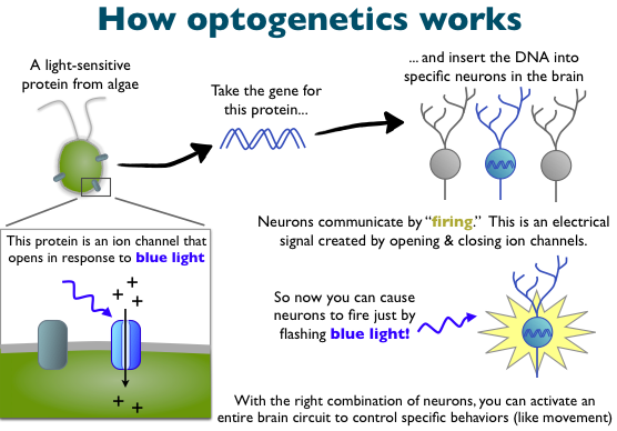
Optogenetics, the process of controlling brain cells using light, could be used to understand and treat brain diseases, including epilepsy and depression.   Previously, scientists relied on fMRI and a wire probe inserted into the brain to switch on cells.

The technique must be refined before it can be used in people or in remote parts of the brain.

Columbia’s Elizabeth Hillman describes the process:   “Instead of activating just one brain cell at a time with a probe, researchers had a way to cause large groups of cells to fire without touching them. You can select that very specific genetic cell type, and you can tell that specific cell type to react when you shine light on it.  First, though, scientists are going to have to overcome some big challenges.  You’re actually altering the genes of the neurons.”  That’s because most neurons don’t normally respond to light.  Genetic material must be added to every brain cell to control it.  Scientists can do that in mice with genetic engineering, but not in people.  Professor Hillman continues: “Another challenge for optogenetics has to do with delivering light to cells deep in the brain. It’s really hard to get light to go deep,and we all know this just from trying to shine a flashlight through our hand.”

Optogenetics is already changing our understanding of epilepsy.

According to Berkeley professor Hillel Adesnik, “Scientists have known for a long time that epileptic seizures occur when brain cells start firing out of control. But they’ve been struggling to understand the role of brain cells called inhibitory neurons, which can reduce firing in other cells. Prior to optogenetics, there was no way to control these neurons and test hypotheses. Now scientists have shown that by altering that activity of inhibitory neurons in mice, it’s possible to start and stop epileptic seizures.”
Categories
BCI Brain Sensors

Microscale torsional muscle system can simulate active neuromuscular system

http://newscenter.lbl.gov/news-releases/2013/12/19/a-micro-muscular-break-through/

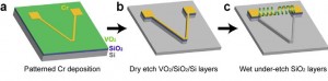
U.S. Department of Energy and Lawrence Berkeley National Laboratory researchers have developed a micro-sized robotic torsional muscle/motor made from vanadium dioxide. For its size, it is a thousand times more powerful than a human muscle, able to catapult objects 50 times heavier than itself over a distance five times its length within 60 milliseconds.

“Multiple micro-muscles can be assembled into a micro-robotic system that simulates an active neuromuscular system,” said Berkely professor Junqiao Wu.  “The naturally combined functions of proximity sensing and torsional motion allow the device to remotely detect a target and respond by reconfiguring itself to a different shape. This simulates living bodies where neurons sense and deliver stimuli to the muscles and the muscles provide motion.”

Categories
AI BCI Brain Crowdfunding

Crowdfunded 3-D augmented reality glasses aim to compete with Google Glass

https://www.spaceglasses.com/

For several months, after a successful Kickstarter campaign, Meta has been developing augmented reality glasses “that combine the power of a laptop and smartphone in a pair of thick Ray-Bans and a small pocket computer.”

The Meta Pro will have an i5 CPU, 4GB of RAM, 128 GB of storage, Wi-Fi 802.11n and Bluetooth 4.0 connectivity. It will cost $3,000, and the company hopes to ship by June.

Meta is hoping to compete with Google Glass, although it is not currently wireless.  The glasses have 15x the display of Google Glass, and runs 3-D instead of 2-D. Its optics are thinner, at 2mm vs 5mm, and its sensors recognize hand gestures, which makes control easier than touching the side of your face.

Categories
BCI Brain

EEG patch monitors attention in students

 http://www.calcalist.co.il/local/articles/0,7340,L-3619747,00.html

http://www.calcalist.co.il/local/articles/0,7340,L-3620186,00.html

Professor Nathan Intrator of Tel Aviv University’s Blavatnik School of Computer Science and Sagol School of Neuroscience, and Guy Levi, Chief Innovation Officer of Israel’s Center for Educational Technology are disrupting education through brain science.

In an attempt to improve learning abilities in children with attention issues, Intrator and Levi attach a patch to student’s foreheads to measure brain activity during lessons.  The learning process can then be adapted to their skills and needs. A student’s lack of attention is identified and better methods or times for delivery of information are suggested.

Through advanced signal processing, Intrator uses a patch with three electrodes to extract information about attention and cognitive strategies.

This less obtrusive approach allows widespread use of EEG in diagnostic studies. Intrator’s current focus includes attention, dementia, and sleep.

Categories
Brain fMRI

fMRI shows emotional reactions in vegetative patients

http://www.plosone.org/article/info:doi/10.1371/journal.pone.0074711

Using fMRI, Tel Aviv University and Sourasky Medical Center’s Haggai Sharon, Yotam Pasternak, Talma Hendler and colleagues have shown that the brains of patients in a vegetative state emotionally react to photographs of people they know, as though they recognize them.

“We showed that patients in a vegetative state can react differently to different stimuli in the environment depending on their emotional value,” said Dr. Sharon. “It’s not a generic thing; it’s personal and autobiographical. We engaged the person, the individual, inside the patient.”

Research focused on the “emotional awareness” of patients in a vegetative state is relatively new. The researchers hope to eventually contribute to improved care and treatment. They are also working with patients in a minimally conscious state to better understand how regions of the brain interact in response to familiar cues.

Categories
Brain

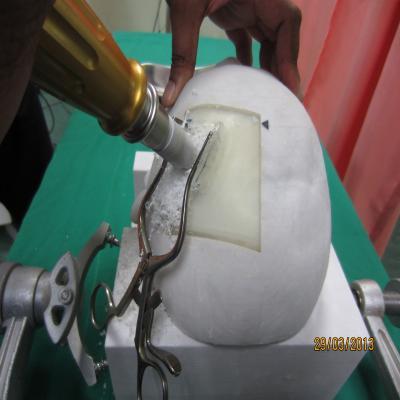
Medical students simulate surgery on 3-D printed brains

http://www.npr.org/blogs/health/2013/12/16/250577798/novice-neurosurgeons-train-on-brains-printed-in-3-d

University of Malaya neurosurgeons use 3-D printers to make realistic skulls and brains for surgical residents.  The models combine different materials to mimic the feel of human bone, membrane and tissue. Each practice patient is made to order from the scans of an actual patient, so students can attempt the same procedures as senior surgeons.

At the University of Florida, neurosurgeons have combined a similar 3-D printed model with a visual simulator similar to fluoroscopy.

Categories
AI BCI Brain

Year end review of “neuromorphic” chip prototypes

http://www.technologyreview.com/featuredstory/522476/thinking-in-silicon/
MIT Technology Review today features an overview of processors that they claim are “about to narrow the gulf between artificial and natural computation—between circuits that crunch through logical operations at blistering speed and a mechanism honed by evolution to process and act on sensory input from the real world.”

Caltech’s Carver Mead pioneered “brain inspired” computing in the 1980’s, based on theoretical math and logic.  ApplySci has featured related research from The University of Zurich/ETH, DARPA, Intel, IBM, Qualcomm, and others, as well as the “deep learning” initiatives of Google and Facebook.  We anticipate and will report on advances in this space in the coming year.

Categories
AI Assistive Technologies BCI Brain

Nerve interface simulates touch in prosthetic hand

http://www.technologyreview.com/news/522086/an-artificial-hand-with-real-feelings/
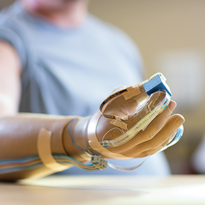
Cleveland Veterans Affairs Medical Center and Case Western Reserve University researchers have developed an interface that can convey a sense of touch from 20 spots on a prosthetic hand. It directly stimulates nerve bundles, known as peripheral nerves, in the arms of patients.   Two people have been fitted with the interface to date. The implants continue to work after 18 months, which is notable because electrical interfaces to nerve tissue can gradually degrade in performance.

According to Case Western Professor Dustin Miller, who is leadning the project:  “The work opens up the possibility that prosthetic limbs could one day provide enduring and nuanced feedback to humans.”

Categories
Brain Seniors

Brain scan for Alzheimer’s diagnosis

http://www.imperial.nhs.uk/aboutus/news/news_042335
Tomorrow, London’s Imperial College will be the first to perform a brain scan which could lead to more accurate diagnosis of Alzheimer’s disease and other dementias.

The test involves giving the patient a small amount of a radiopharmaceutical which binds to amyloid plaques, showing them in a brain scan. Amyloid plaques in the brain are one of the hallmarks of Alzheimer’s disease, so a negative scan effectively excludes a diagnosis of the condition.

This is the first time that clinicians have been able to see these plaques while a patient is alive, marking a significant breakthrough in the diagnosis of dementia. The presence of plaques in the new scan will also help to ensure that patients with memory problems who take part in clinical trials for amyloid-targeting drugs are appropriately selected.

Until now, Alzheimer’s disease could only be diagnosed definitively via brain autopsy.

Categories
Brain fMRI

Imaging technology identifies signs of chronic brain injury in living football players

http://www.technologyreview.com/news/522126/identifying-signs-of-chronic-brain-injury-in-living-football-players/

Until now, chronic traumatic encephalopathy, caused by repetive head injury, could only be identified after a victim died.   CTE is linked to depression, dementia, and memory loss.

A new imaging method, developed by Pittsburgh Steelers physician Julian Bailes and UCLA researchers, can for the first time spot signs of the condition in the living brain.  It could help players avoid the degenerative condition by limiting their exposure, and it may help scientists develop better protective gear and treatments.

The technology is based on a positron emission tomography scans. UCLA researchers developed a radioactive compound that can be injected intravenously. The compound circulates through the bloodstream and into the brain, where it gloms onto tau proteins, which can then be measured in a PET scanner. The test takes about an hour.

The radioactive compound also sticks to amyloid proteins. Aggregations of both amyloid and tau are considered culprits in Alzheimer’s disease, whereas tau is the main indicator for CTE. Bailes and colleagues say the regions of the brain that are highlighted in PET scans of patients with Alzheimer’s differ from the scans of patients with CTE.

In related research, Israeli company ElMindA is developing non-invasive BNA (brain network activation) technology.  Patients sit at a computer for 15 to 30 minutes, performing a specific task many times while the device maps network activation points in the brain. The repetition allows the device to sift out brain activity unrelated to the task.  The result is a three-dimensional image of nerve cell connectivity and synchronization that is highly sensitive, specific and reproducible.  The tool is sensitive enough to show subtle differences in the severity of the condition from one day to another, according to the company. They claim that it can also optimize drug dosing by monitoring the changes in brain network activities as the drug takes effect.

Categories
Assistive Technologies BCI Brain Sensors

Nerve impulse sensor exoskeleton assists paraplegics, Parkinson’s, stroke patients

http://www.dw.de/standing-again-with-nerve-controlled-robotics/a-17280419

Professor Thomas Schildhauer leads a team at Bergmannsheil University Clinic’s “Center for Neuro-Robotic Mobility Training”  that uses nerve impulse sensors to help patients walk again.  A robotic exoskeleton with sensors affixed to the hips and legs gives paraplegics, Parkinson’s and stroke patients a sense of stability during ambulatory exercises. The robot suit contains numerous sensors that recognize nerve impulses as they flash across the skin. Via a small motor, the suit converts those impulses into motion.

“The brain sends a signal out that typically arrives at the muscle via nerve systems,” said Schildhauer.  For patients capable of some movement, “Small impulses can still be discovered in the muscles. And they can be measured and recorded on the skin. That signal is then amplified in the robot and moves the motors of the exoskeleton.”

Such robot-supported training, Schildhauer says, “seems to build up and expand the remaining muscle functions, and the brain structures, too, that haven’t been used for a long time.” Movement patterns, he added, are then re-trained. “It seems to cause the patient to fall back into many of the old, usual cycles of movement, and results in them being able to walk again.”

Categories
Brain Sensors

Molecular sensor for early detection of Multiple Sclerosis

http://onlinelibrary.wiley.com/doi/10.1002/ana.24078/abstract

UCSF’s Gladstone Institute researchers have created a molecular sensor that can detect MS at its earliest stages — before the onset of physical signs. Professor Katerina Akassoglou’s study revealed in animal models that the heightened activity of a protein called thrombin in the brain could serve as an early indicator of MS. By developing a fluorescently labeled probe specifically designed to track thrombin, the team found that active thrombin could be detected at the earliest phases of MS, and that this active thrombin correlates with disease severity.

In laboratory experiments on mice modified to mimic the signs of MS, the team employed an Activatable Cell-Penetrating Peptide, a molecular probe that delivers fluorescent agents to a region of interest. For this study, they developed a thrombin-specific probe that could track thrombin activity in mice as the disease progressed. They then analyzed where, and at what stage of disease, thrombin activity was found.

“We detected heightened thrombin activity at specific disease ‘hot-spots,’ regions where neuronal damage developed over time,” said Gladstone scientist Dimitrios Davalos, “And when we compared these results to those of a separate, healthy control group of mice, we saw that thrombin activity in the control group was wholly absent.”