
Categories
Autism Brain Eyes Machine Learning Parkinson's

Eye tracking data helps diagnose autism, ADHD, Parkinson’s

http://www.scientificamerican.com/article.cfm?id=eye-tracking-software-may-reveal-autism-and-other-brain-disorders

USC’s Laurent Itti and researchers from Queen’s University in Ontario have created a data heavy, low cost method of identifying brain disorders through eye tracking.  Subjects watch a video for 15 minutes while their eye movements are recorded. An enormous amount of data is generated as the average person makes three to five saccadic eye movements per second.  Itti’s team uses advanced machine learning algorithms to enable a computer to recognize patterns without explicit human instruction.

The proof of concept study found that the algorithm could classify mental disorders through eye movement patterns.  Parkinson’s patients were identified with nearly 90 percent accuracy.  Children with ADHD or fetal alcohol spectrum disorder were identified with 77 percent accuracy.  “This is very different from what people have done before. We’re trying to have completely automated interpretation of the eye movement data,” said Itti.
Categories
Brain fMRI Machine Learning

fMRI and machine learning identify emotions

http://www.plosone.org/article/info%3Adoi%2F10.1371%2Fjournal.pone.0066032

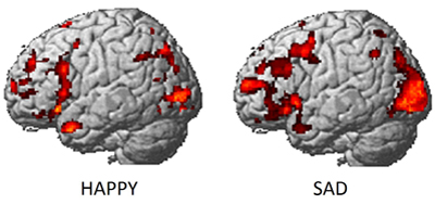
Carnegie Mellon researchers have developed “a new method with the potential to identify emotions without relying on people’s ability to self-report” using a combination of fMRI and machine learning.

They recruited 10 actors from the university’s drama school to act out emotions including anger, happiness, pride and shame, while inside an fMRI scanner,  multiple times in random order.  To ensure that researchers were able to measure actual emotions and not just the acting out of emotions, participants viewed emotion-eliciting images while undergoing fMRI scans.

The findings illustrate how the brain categorizes feelings, giving researchers the first reliable process to analyze emotions.  Emotion research has been difficult because of a lack of reliable evaluation methods, caused by a subject’s reluctance to honestly report feelings and the potential of emotional responses not being consciously experienced. Researchers plan to apply this new identification method to a number of problems, including identifying emotions that individuals are attempting to suppress and multiple emotions experienced simultaneously.

Categories
Brain

Highly detailed 3-D brain image unveiled

http://spectrum.ieee.org/tech-talk/biomedical/imaging/bigbrain-project-makes-terabyte-map-of-a-human-brain

Canadian and German neuroscientists have unveiled the most detailed 3-D image of the human brain to date.  It reveals structures as tiny as 20 microns, 50 times smaller than those created using the best MRI technology.  The image, created as part of a project called the BigBrain, is part of a larger effort to create a high-resolution computer model of the human brain that can serve as a reference point for future studies.  Data from other studies can be combined with this model to allow scientists to link brain function to specific groups of nerve cells.  The information can be used to test theories about brain activity and lead to treatments for diseases.  Until now, brain scans used MRI and PET technology, which can only capture structures as small as a millimeter. This had not been enough to understand what happens when a person gets Alzheimer’s disease or epilepsy.

Categories
AI BCI Brain

Low cost GPU based neural network simulates the brain

http://stanford.edu/~acoates/papers/CoatesHuvalWangWuNgCatanzaro_icml2013.pdf

In a new paper, Stanford’s Andrew Ng describes how to use graphics microprocessors to build a $20,000 computerized brain that is similar to the cat-detector he developed with Google last year for $1M.

To test his hypothesis about GPU-driven Deep Learning, he also built a larger version of the platform for $100,000.  It utilized 64 Nvidia GTX 680 GPUs on 16 computers. It was able to accomplish the same cat-spotting tasks as the Google system, which needed 1,000 computers to operate.  That system, modeling the activities and processes of the human brain, was able to learn what a cat looks like, and then translate that knowledge into spotting different cats across multiple YouTube videos.

Categories
Brain mHealth Monitoring Sensors

Sports sensors warn of head injury

http://www.nytimes.com/2013/06/16/business/a-wearable-alert-to-head-injuries-in-sports.html?_r=0
The New York Times reports on the growing trend of sensor based protection/early warning systems for athletes. The devices, packed with sensors and microprocessors, register a blow to a player’s skull and immediately signal the news by blinking brightly, or by sending a wireless alert.  Algorithms evaluate the impact and determine severity.


Categories
Brain

Brain scans and depression treatment outcomes

http://archpsyc.jamanetwork.com/article.aspx?articleid=1696349

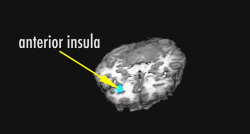
In an NIH funded clinical trial, researchers at Emory University have discovered that specific patterns of brain activity may indicate whether a depressed patient will or will not respond to treatment with medication or psychotherapy.

Professor Helen Mayberg, MD and colleagues used PET scans to measure brain glucose metabolism, an important index of brain functioning to test this hypothesis.  Participants in the trial were randomly assigned to receive a 12-week course of either the SSRI medication escitalopram or cognitive behavior therapy after first undergoing a pretreatment PET scan.  The team found that activity in one particular region of the brain, the anterior insula, could discriminate patients who recovered from those who were non-responders to the treatment assigned. Specifically, patients with low activity in the insula showed remission with CBT, but poor response to medication; patients with high activity in the insula did well with medication, and poorly with CBT.

Categories
AI BCI Brain

Cornell robots anticipate human actions

http://news.cornell.edu/stories/2013/04/think-ahead-robots-anticipate-human-actions
Cornell University researchers have programmed a PR-2 robot to not only carry out everyday tasks, but to anticipate human behavior and adjust its actions.

From a database of 120 3-D videos of people performing common household activities, the robot has been trained to identify human activities by tracking the movements of the body – reduced to a symbolic skeleton for easy calculation – breaking them down into sub-activities like reaching, carrying, pouring or drinking, and to associate the activities with objects.

Observing a new scene with its Microsoft Kinnect 3-D camera, the robot identifies the activities it sees, considers what uses are possible with the objects in the scene and how those uses fit with the activities; it then generates a set of possible continuations into the future – such as eating, drinking, cleaning, putting away – and finally chooses the most probable. As the action continues, it constantly updates and refines its predictions.

Categories
Brain fMRI

Advanced imaging modalities map neural connections

http://spectrum.ieee.org/biomedical/imaging/a-wiring-diagram-of-the-brain

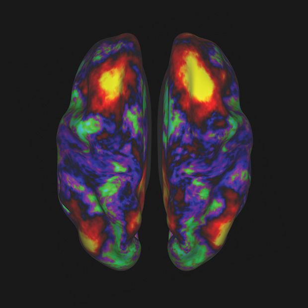
Images of 68 brains from the Human Connectome Project recently became available. The process was powered by highly advanced brain scanning hardware and state of the art image processing and analysis software.

To provide multiple perspectives on each brain, researchers employed several methods:

1. MRI scans provided basic structural images of the brain, providing very high resolution images of the convoluted folds of the cerebral cortex.

2. fMRI scans detected blood flow throughout the brain and showed brain activity for subjects both at rest and engaged in seven different tasks (including language, working memory, and gambling exercises).

3. Diffusion MRI tracked the movement of water molecules within brain fibers. Because water diffuses more rapidly along the length of the fibers that connect neurons than across them, this technique allows researchers to directly trace connections between sections of the brain.

Each imaging modality has its limitations, so combining them gives neuroscientists the best view of how the brain works.  The data was purged of noise and artifacts, and then organized into a database.

Categories
Brain

Multitasking neurons enhance brain’s computational power

http://web.mit.edu/newsoffice/2013/complex-brain-function-depends-on-flexibility-0519.html

Many neurons, especially in brain regions that perform sophisticated functions such as thinking and planning, react differently to a wide variety of stimulation.

“We started noticing early on that there are a whole bunch of neurons in the prefrontal cortex that can’t be classified in the traditional way of one message per neuron,” said Professor Earl Miller of MIT’s Picower Institute.

Miller and colleagues report that these neurons are essential for complex cognitive tasks, such as learning new behavior.  Professor Stefano Fusi at Columbia University developed a computer model showing that without these neurons, the brain can learn only a handful of behavioral tasks.

Categories
Brain Seniors Stroke

Hyperbaric oxygen treatment for brain injuries

http://www.assafh.org/sites/en/Pages/brain-injuries.aspx

Tel Aviv University and Assaf Harofeh Medical Center researchers are treating stroke patients with hyperbaric oxygen therapy (HBOT), high-pressure chambers where oxygen-rich air increases oxygen levels in the body by a factor of ten.  Their goal is to reinvigorate dormant neurons and improve patients’ motor function, memory and other abilities that current therapies do not address.

The researchers are studying the potential benefits of HBOT for traumatic brain injury, and as an anti-aging therapy, applicable to Alzheimer’s and vascular dementia patients.

Categories
Brain Sensors

Wireless detection of brain trauma

http://health.universityofcalifornia.edu/2013/05/14/wireless-signals-could-transform-brain-trauma-diagnostics/
New technology developed at UC Berkeley uses wireless signals to provide real-time, noninvasive diagnoses of brain swelling or bleeding. The device analyzes data from low-energy electromagnetic waves, similar to the kind used to transmit radio and mobile signals.  It is sensitive enough to distinguish between a normal brain and a diseased brain with one single noncontact set of measurements.

Categories
BCI Brain EEG Machine Learning Monitoring

Skin mounted electrode arrays measure neural signals

http://coleman.ucsd.edu/lab-research/

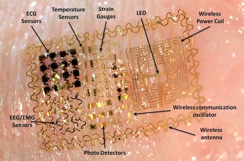
Professor Todd Coleman of UCSD is developing foldable, stretchable electrode arrays that can non-invasively measure neural signals. They can also provide more in-depth analysis by including thermal sensors to monitor skin temperature and light detectors to analyze blood oxygen levels.  The device is powered by micro solar panels and uses antennae to wirelessly transmit or receive data.  Professor Coleman wants to use the device on premature babies to monitor their mental state and detect the onset of seizures that can lead to brain development problems such as epilepsy.