Categories
Brain fMRI Neural Computation

Computational modeling and fMRI show the roles of brain regions in behavioral control

https://www.cell.com/neuron/abstract/S0896-6273(13)01125-2

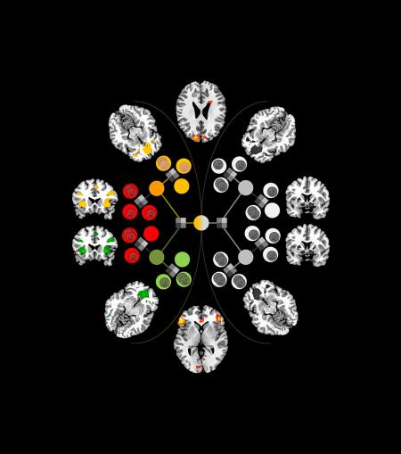
John O’Doherty of the Caltech Brain Imaging Center has pinpointed areas of the brain—the inferior lateral prefrontal cortex and frontopolar cortex—that seem to serve as the “arbitrator” between model-based and model-free decision-making systems, weighing the reliability of the predictions each makes and then allocating control accordingly.  Professor O’Doherty believes that this can lead to better treatments for brain disorders, such as drug addiction, and psychiatric disorders, such as obsessive-compulsive disorder. These disorders, which involve repetitive behaviors, may be driven in part by malfunctions in the degree to which behavior is controlled by the habitual system versus the goal-directed system.

Categories
Assistive Technologies Brain Heart Stroke

Non-invasive, nanoparticle method for identifying atherosclerosis plaques

http://pubs.acs.org/doi/abs/10.1021/nl404816m

Case Western‘s Michael Bruckman and colleagues have developed a multifunctional nanoparticle that pinpoints blood vessel plaques caused by atherosclerosis using MRI.  The goal is to create a non-invasive method of identifying heart attack and stroke causing plaques vulnerable to rupture, in time for treatment.

Currently doctors can only identify narrowing blood vessels caused by plaque accumulation via incision and the insertion of a catheter inside a blood vessel in the arm, groin or neck. The catheter emits a dye that enables X-rays to show the narrowing.

The researchers found that a nanoparticle built from a rod-shaped virus, commonly found on tobacco, locates and illuminates plaque in arteries more effectively, with a fraction of the dye.  The tailored nanoparticles target plaque biomarkers, opening the possibility that particles can be programmed to identify vulnerable plaques from stable.  Untargeted dyes alone cannot accomplish this.

Categories
Prosthetics Robotics Sensors

Trial: Improved sense of touch and control in prosthetic hand

http://stm.sciencemag.org/content/6/222/222ra19

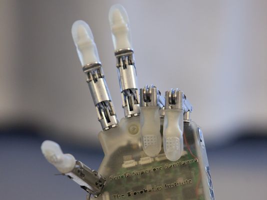
In the ongoing effort to improve the dexterity of prosthetics, a recent trial showed an improved sense of touch and control over a prosthetic hand.  EPFL professor Silvestro Micera and colleagues surgically attached electrodes from a robotic hand to a volunteer’s median and ulnar nerves. Those nerves carry sensations that correspond with the volunteer’s index finger and thumb, and with his pinky finger and the edge of his hand, respectively. The volunteer controlled the prosthetic with small muscle movements detected by sEMG, a non-invasive method that measures electrical signals through the skin.

Over seven days, the volunteer was asked to grasp something with a light grip, a medium grip, and a hard grip, and to evaluate the shape and stiffness of three kinds of objects. During 710 tests, he wore a blindfold and earphones so that he could not use his vision or sound to guide the prosthetic. The researchers also sometimes turned off the sensory feedback to test whether he was using time to modulate his grip.

The subject was able to complete the requested tasks with his prosthetic thumb and index finger 67 percent of the time the first day and 93 percent of the time by the seventh day of the experiment. His pinky finger was harder to control: he was only able to accomplish the requested grip 83 percent of the time. In both the grip strength tests and in detecting the stiffness of objects, the volunteer made mistakes with the medium setting and object, but he never confused the softest and hardest objects. The ability to modulate his grip strength is this study’s main progress over previous work by the same group.

Categories
Cancer Ultrasound

Ultrasound combined with contrasting agent for radiation-free tumor detection

http://www.plosone.org/article/info%3Adoi%2F10.1371%2Fjournal.pone.0086642

University of North Carolina Professor Nancy Klauber-Demore has improved the resolution and tumor-detecting ability of ultrasound scans.

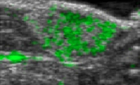
Combining ultrasound with a contrast agent composed of tiny bubbles that pair with an antibody that many cancer cells produce at higher levels than do normal cells, Klauber-Demore was able to visualize lesions created by angiosarcoma.  By binding to the protein SFRP2, the contrast agent may help distinguish malignant from benign masses found on imaging.  Since SFRP2 is expressed in many cancers – including breast, colon, pancreas, ovarian, and kidney tumors – the technique could potentially be useful for a broad range of cancer types.  As the level of SFRP2 in tumors increases as tumors develop, the team will also investigate whether the technique can be used to track tumor growth.