Categories
Brain Cancer Ultrasound

Ultrasound penetrates blood-brain barrier to treat brain tumor

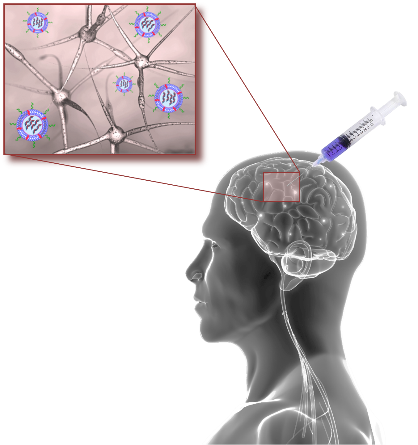
Todd Mainprize at Sunnybrook Hospital has, for the first time,  delivered chemotherapy directly to a brain tumor, by breaking through the blood-brain barrier using tightly focused ultrasound.

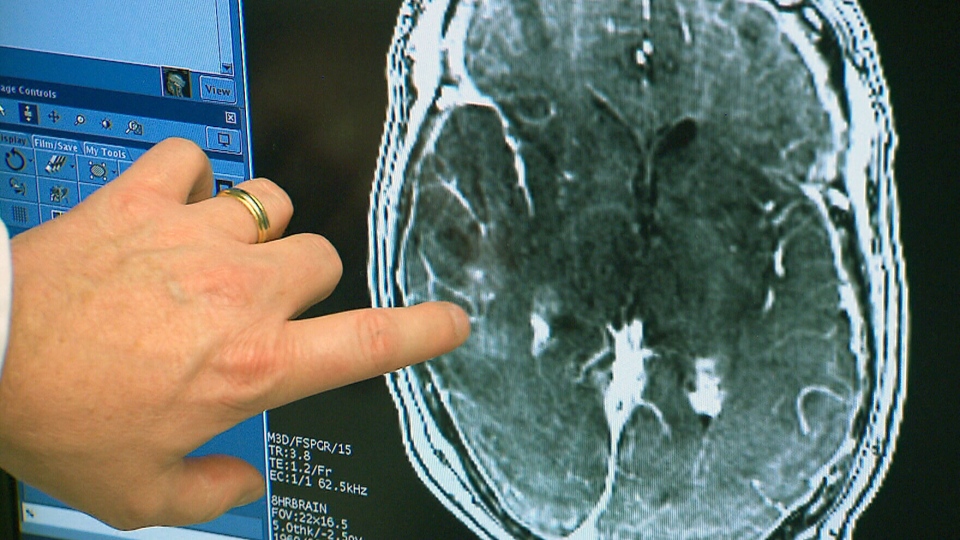
The patient’s bloodstream was  infused with a chemotherapy drug, as well as microscopic bubbles, which are smaller than red blood cells and can pass freely through blood. MRI-guided, low intensity sound waves targeted blood vessels in the blood-brain barrier, near the tumor site. The ultrasound waves vibrated the microbubbles,  loosening the tight cell junctions of the blood-brain barrier. The loosened junctions allowed the chemotherapy drug to flow past the barrier and deposit within the targeted tumor site.

This breakthrough could also lead to new treatments for brain diseases such as Parkinson’s and Alzheimer’s.

Click to view Sunnybrook Hospital video.

NEUROTECH SAN FRANCISCO – APRIIL 6, 2016 @ THE MISSION BAY CONFERENCE CENTER
Categories
Cancer Sensors

Remotely controlled capsule endoscope captures lower GI images

A new type of capsule endoscope may improve cancer diagnostics, providing comprehensive, non-invasive imaging, including lower GI images.

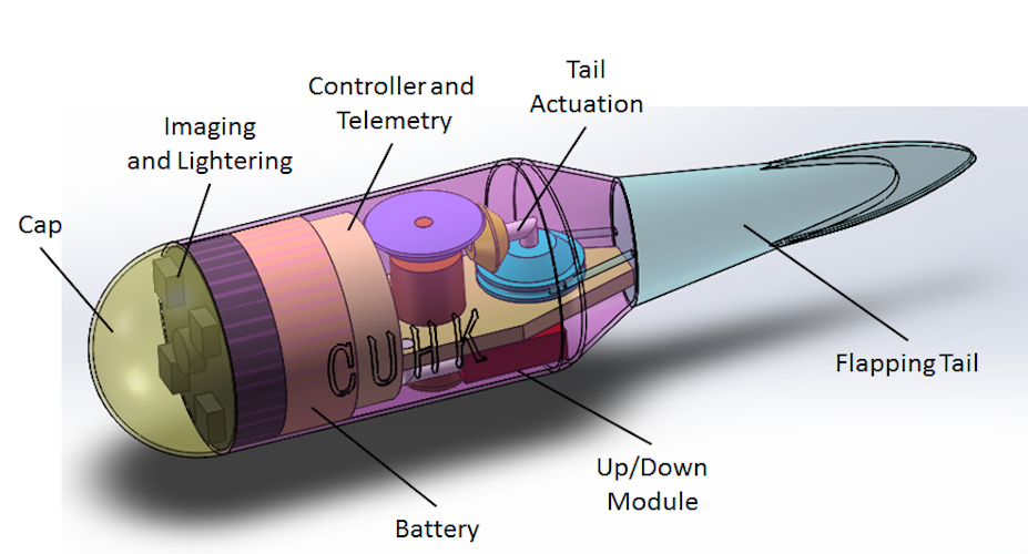
The 3D printed Tadpole Endoscope (TE) has a soft tail that allows it to be  remotely guided around the stomach.  The technology was developed by Yong ZHONG, Ruxu DU and Prof Phillip W Y CHIU of the Chinese University of Hong Kong.

The TE is activated immediately after being swallowed. Once it reaches the stomach, a doctor can guide the device to gather images. By adjusting a patient’s posture, the whole stomach can be viewed. The TE moves into the lower GI tract via natural peristalsis. The patient is sent home, wearing a sensor patch, to record  the lower GI images, which are transmitted to the doctor.

Currently, esophagus and stomach cancer can be diagnosed using gastroscopy; intestinal cancer can be diagnosed using capsule endoscopy; and colorectal cancer can be diagnosed using colonoscopy.

Click to view the Chinese University of Hong Kong video.

Categories
3-D Printing Cancer

Cancer patient receives 3D printed rib cage

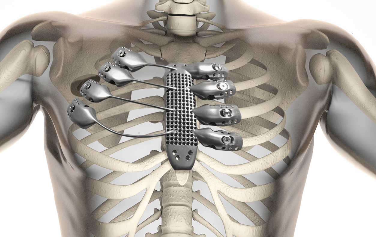
For the first time, a chest wall sarcoma patient has received a  fully customized 3d printed sternum and rib cage portion, created using high resolution CT data.

This part of the chest is difficult to recreate with traditional prosthetics.  Thoracic surgeons typically use flat and plate implants for the chest, which can loosen over time and increase complications.  Rapidly prototyped 3D printed ribs may become the future standard.

View CSIRO video here.

Categories
Cancer Sensors

Biochemical sensor helps refine cancer treatment

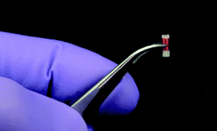
MIT professor Michael Cima is developing a tiny biochemical sensor that can be implanted in cancerous tissue during a biopsy.  It wirelessly sends  biomarker data to an external device,  allowing doctors to monitor progress, and adjust dosages or switch therapies accordingly.

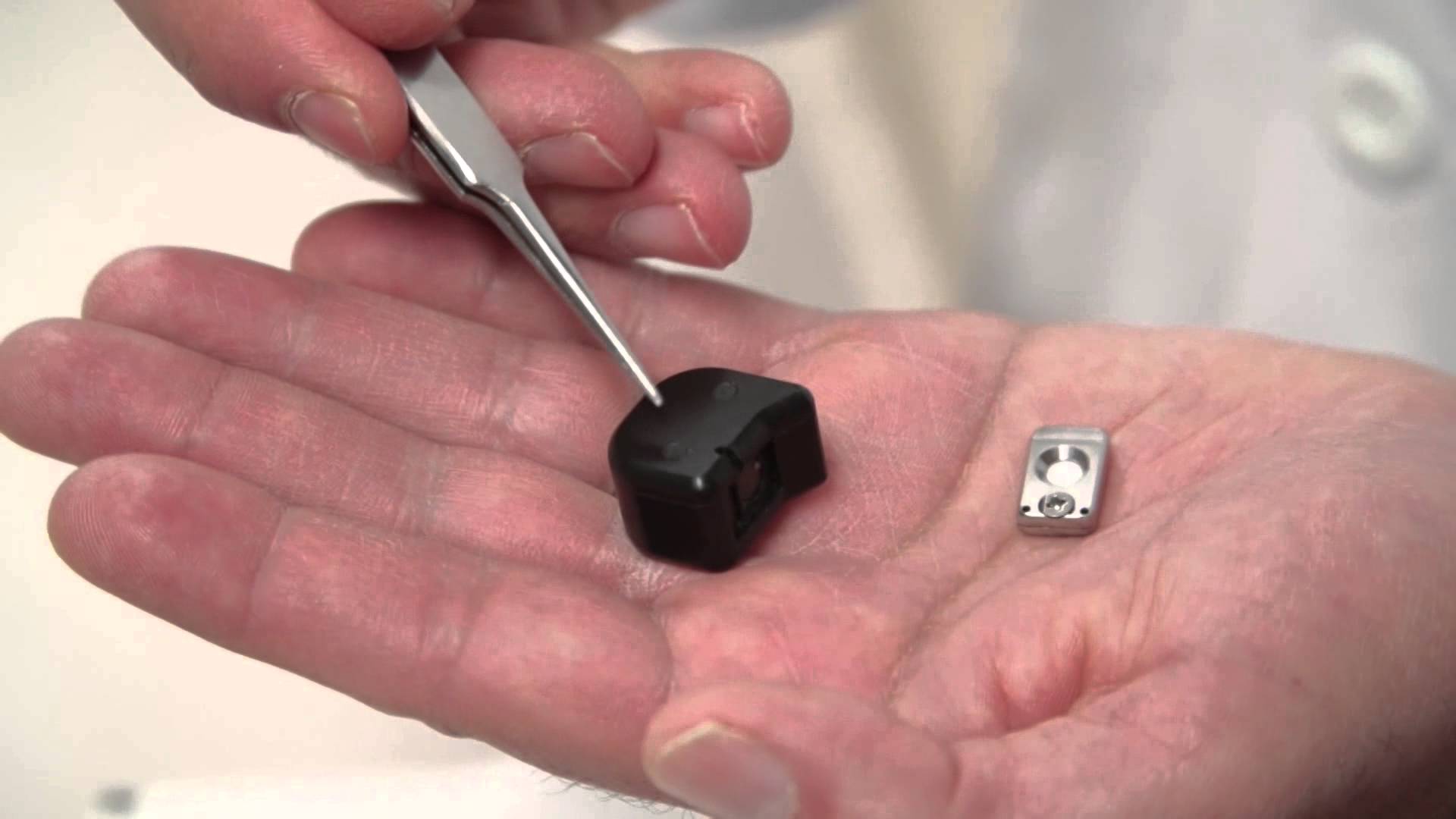
The sensor fits into the tip of a biopsy needle. It contains 10 microliters of chemical contrast agents typically used for MRI, and an on-board circuit to communicate with the external reader. Real-time, on-demand data concerning two biomarkers linked to a tumor’s response to treatment, pH and dissolved oxygen, is provided.

The goal is to make treatments more targeted and precise, to improve their efficacy and reduce side effects.

Categories
Cancer Sensors

Sensor chip for prostate cancer diagnosis

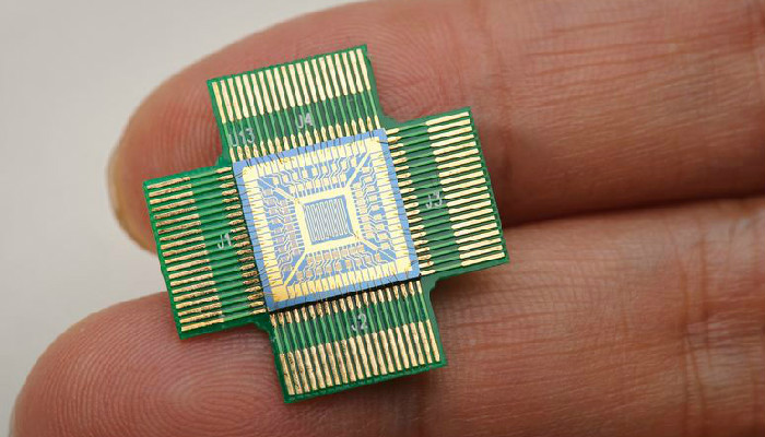
University of Birmingham researchers are developing a sensor chip that they believe can improve the accuracy of prostate cancer diagnosis.

Prostate cancer is normally diagnosed by tests that rely on antibodies, making them vulnerable to degeneration by environmental changes. They are known to give false positive readings at a high rate.

The sensor chip works by identifying glycoprotein molecules frequently used as biomarkers for diseases, including prostate cancer.  Detection techniques have focused on the protein of the molecule, which does not always change when the body is affected by disease.

The new chip uses  synthetic receptors along a 2D surface to identify specific, targeted glycoprotein molecules that are differentiated by their modified carbohydrate chains.  This shows subtle differences in healthy and diseased patients.

Categories
Cancer Pain

Remote controlled nanowire drug delivery

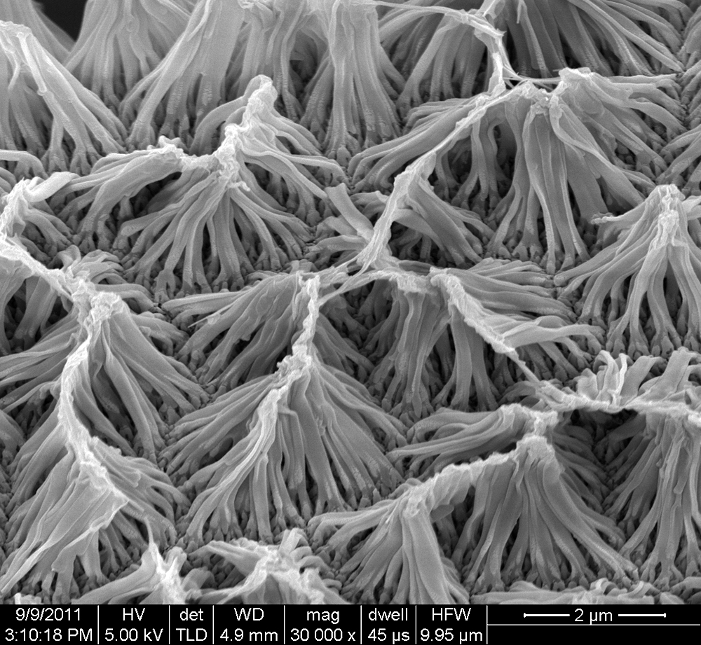
Purdue researchers have created an implantable drug-delivery system using nanowires that can be controlled wirelessly.

The nanowires respond to an electromagnetic field generated by a device used to control the release of a preloaded drug. Tubes and wires required by other implantable devices are eliminated, minimizing the risk of infection and  complications.

According to lead author Richard Borgens: “This tool allows us to apply drugs as needed directly to the site of injury, which could have broad medical applications, The technology is in the early stages of testing, but it is our hope that this could one day be used to deliver drugs directly to spinal cord injuries, ulcerations, deep bone injuries or tumors, and avoid the terrible side effects of systemic treatment with steroids or chemotherapy.”

2 DAYS UNTIL WEARABLE TECH + DIGITAL HEALTH NYC 2015 – JUNE 30 @ NEW YORK ACADEMY OF SCIENCES.  REGISTER HERE.

Categories
Cancer Sensors

Chip detects cancer from a drop of blood

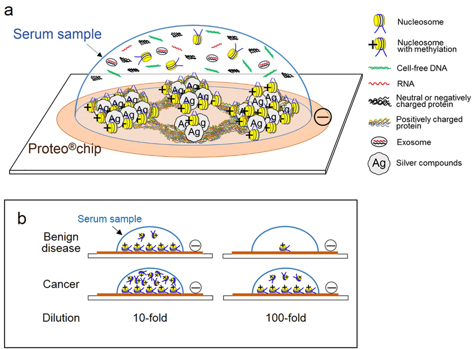
Showa University and My Tech have developed Proteo, a silver nanoscale chip that  they claim detects most types of cancer from a drop of blood in three minutes.   It functions by attracting a faintly luminous substance found in cancer patients, beginning at a very early stage.

They have only studied 20 patients, but diagnosed whether a tumor is benign or malignant with total accuracy.

The researchers are accumulating data from various types of cancers, and hope to make the blood test available next year. They believe that the preliminary data suggests that the device could be more effective than existing blood tests.

Showa’s Hiroaki Ito and MyTech’s Yuki Hasegawa said that the accuracy derives from the chip attracting certain nucleosomes, which are prevalent in all types of cancer.  The nucleosome emits light, the intensity of which increases as more of the substance accumulates.  This is seen under a fluorescent microscope, providing the ability to  identify visually if the blood is from an individual with cancer.

WEARABLE TECH + DIGITAL HEALTH NYC 2015 – JUNE 30 @ NEW YORK ACADEMY OF SCIENCES.  REGISTER HERE.

Categories
Cancer Wearables

Wearable tracks breast cancer side effects, sleep, mood, activity

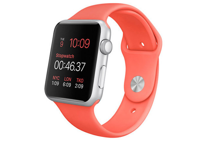
A pilot study exploring  the use of wearables in breast cancer is underway.  Polaris Health Directions and  the MD Anderson Cancer Center are using the Apple Watch to  track multiple factors, increase engagement, and provide immediate feedback and interventions.

Side effects, sleep, activity levels and mood will be monitored, and combined with electronic health records and population data.

According to Polaris,  “the patient  will learn through self-discovery  how to modify  behavior, and if she finds herself in a high stress situation, our on-demand support mechanisms can provide instant assistance.”

WEARABLE TECH + DIGITAL HEALTH NYC 2015 – JUNE 30 @ NEW YORK ACADEMY OF SCIENCES.  REGISTER HERE.

Categories
Cancer Seniors

Human cell sensor detects chemical changes early

University of Rochester‘s Spencer Rosero is developing a human cell sensor that, when implanted,  detects subtle biological changes to provide advanced warning of health issues.

The cells are engineered to detect specific chemical changes. When a variation is discovered, the cells respond, and a fluorescent light glows. The sensor’s camera  enables patients and doctors to see the glowing light in real time on a computer or device.  Placement of the sensor (underneath the skin) depends on what is being tracked.

Chemotherapy patients will be the first users.  Toxic side effects will be detected before they happen, to help physicians optimize treatment.

WEARABLE TECH + DIGITAL HEALTH NYC 2015 – JUNE 30 @ NEW YORK ACADEMY OF SCIENCES.  REGISTER HERE.

Categories
Cancer Personalized Medicine

Implant tests cancer drugs to optimize treatment

MIT’s Oliver JonasRobert Langer, and colleagues have developed an implantable device that allows doctors to test cancer drugs in patients before prescribing chemotherapy.

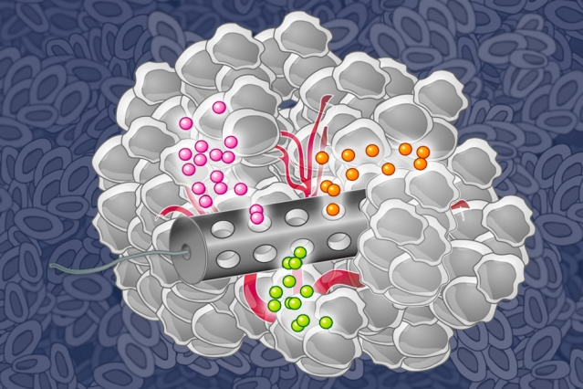
The tiny device can carry small doses of 30 drugs. After implanting it in a tumor and allowing the drugs to diffuse into the tissue, researchers can measure how effectively each destroys a patient’s cancer cells.  The guesswork involved in choosing treatments can be minimized.

Most cancer drugs work by damaging DNA or interfering with cell function. Scientists have developed more targeted drugs, designed to kill tumor cells that carry a specific genetic mutation. However, it is usually difficult to predict whether a particular drug will be effective in an individual patient.

Doctors sometimes extract tumor cells, grow them in a lab, and treat them with different drugs to determine which are most effective.  This process removes the cells from their natural environment, which can play an important role in a tumor’s response to drug treatment.

According to Jonas, the implant  “essentially puts the lab into the patient.  It’s safe, and sensitivity testing can be done in the native microenvironment.”

WEARABLE TECH + DIGITAL HEALTH NYC 2015 – JUNE 30 @ NEW YORK ACADEMY OF SCIENCES.  REGISTER HERE.

Categories
Brain Cancer Nanoparticles

Nanoparticle treatment targets brain tumors

Tel Aviv University Professor Dan Peer is developing a nanoparticle-based process to target glioblastoma cells, previously considered untreatable.

Nanoparticles were injected into tumors, acting as the drug delivery system. Nucleic acid, with interference RNAs, attached to receptors  expressed specifically on glioma cells, and stopped the activity of a key protein that regulates the rapid reproduction of the cancer cells.

The therapy was tested in mouse models affected with gliomas and control groups treated with standard chemotherapy. The results were, according to the researchers, “astonishing.”

Wearable Tech + Digital Health NYC 2015 – June 30 @ New York Academy of Sciences

Categories
Cancer Sensors

Breath test for head and neck cancer

EPFL researchers have developed a sensor that can identify the presence of a head and neck cancer through breath analysis.

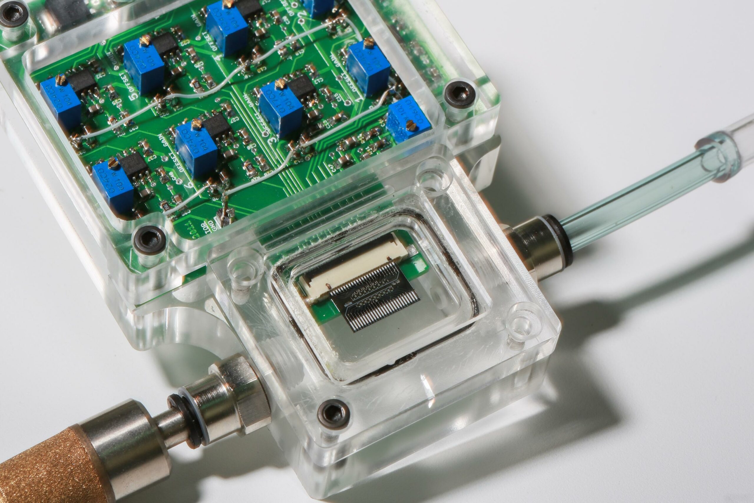
Nico de Rooij‘s micro-sensors  detect volatile organic compounds which vary in  presence and concentration depending on one’s health.

The sensor includes a silicon disk covered by a polymer and suspended by four tiny “bridges” with integrated piezoresistors. When exposed to a gas, the polymer absorbs certain molecules and the disk changes shape. This deformation is detected by the piezoresistive bridges, which emit an electrical signal, determining the signature of the gas and its concentration. Different polymers must be used on each sensor  to obtain an overview of the gas composition.

According to de Rooij, “There are already methods for detecting molecules called ‘electronic noses’ on the market. But they have a hard time analysing very complex gases like human breath, Humidity in particular can disrupt the reading, leading to false positives or false negatives.” The new sensors are extremely accurate.

Wearable Tech + Digital Health NYC 2015 – June 30 @ New York Academy of Sciences