
Categories
Eyes Sensors

Miniature 3D sensor maps surgical field; scope small enough for use in neurosurgery

http://www.visionsense.com/hp.html

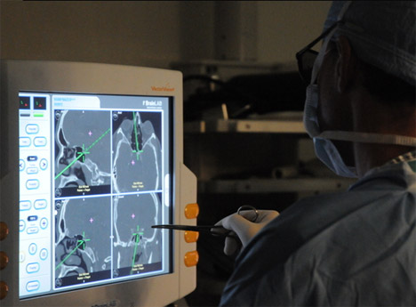
Visionsense’s miniature 3D sensor optically maps the surgical field, based on (and imitating) the eye of a bee. A single sensor is divided into hundreds of thousands of tiny “eyes” looking in different directions, using an array of micron-sized elements.  The elemental information is translated into left and right eye images, resulting in a clear stereoscopic view,  bypassing the diffraction limit that restricts current miniature cameras.

In neurosurgery, scopes must be very small in diameter to pass through narrow ports, such as the nose.  Most 3D scopes rely on two optical channels, each containing a single sensor. Each sensor collects two separate images that are combined to give the appearance of three dimensions as a user looks at the screen – mirroring the sight of the human eye.   Using their single sensor system, Visionsense has developed a high quality image producing scope that is small enough to operate on the brain.

Categories
Diabetes Heart Sensors

Piezoelectric nanoribbons power pacemakers

http://news.cnet.com/8301-11386_3-57617483-76/nanoribbons-let-beating-hearts-power-their-own-pacemakers/

Scientists are studying various steady energy source alternatives for small biomedical sensors, including piezoelectric power.

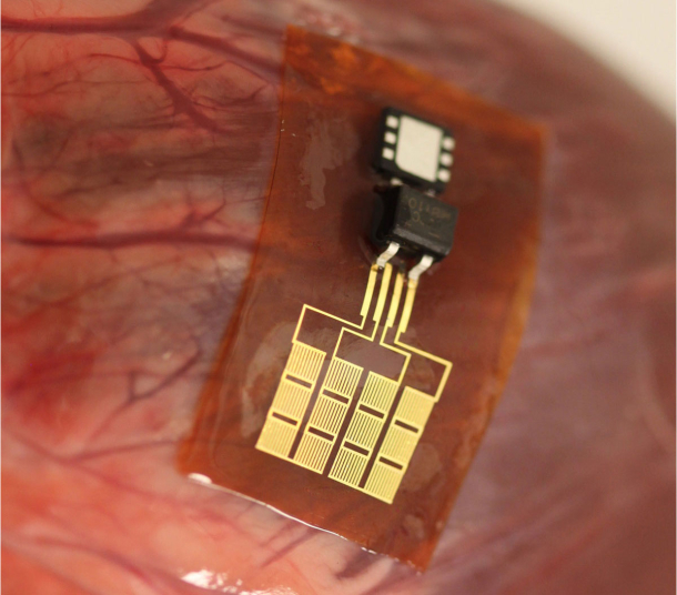
University of Illinois researchers have attached small, flexible strips (piezoelectric nanoribbons) to internal organs of animals, and harvested energy from their movement  to power pacemakers and other medical devices.  Current practice depends on hard-to-change batteries.

This is a minimally invasive power source, which in the future can be activated by muscle activity other than the heart muscle, with potential implications for glucose pumps and other devices.  The researchers claim that their system is a  “complete, flexible, and integrated system that is capable of harvesting and storing energy from the natural contractile and relaxation motions of the heart, lung, and diaphragm at levels that meet requirements for practical applications.”

Categories
Brain Eyes

Brain processes images viewed for 13 milliseconds

http://web.mit.edu/newsoffice/2014/in-the-blink-of-an-eye-0116.html

MIT professor Mary Potter has published a study showing that the human brain is capable of processing images viewed through the eyes for 13 milliseconds. This is significantly faster than the 100 milliseconds reported in earlier research.

The study offers evidence that “feedforward processing” — the flow of information in only one direction, from retina through visual processing centers in the brain — is enough for the brain to identify concepts without having to do any further feedback processing.  It also suggests that while the images are seen for only 13 milliseconds before the next image appears, part of the brain continues to process those images for longer than that.

The researchers are now investigating how long visual information presented so briefly can be held in the brain. They are also scanning subjects’ brains with a magnetoencephalography (MEG) scanner during the task to see what brain regions are active when a person successfully completes the identification task.

Categories
Assistive Technologies BCI Brain

Minimally invasive multi-channel control system for prosthetics tested

http://www.multivu.com/mnr/65112-alfred-mann-foundation-u-s-marine-subject-fda-study-for-imes-system
The Alfred Mann Foundation‘s first subject, a U.S. Marine, will receive its IMES System (implantable myoelectric sensor).   The experimental system could be the first minimally invasive, intuitive, multi-channel control system for prosthetics, intended for long term use. It is being studied under the Investigational Device Exemption regulations of the U.S. Food and Drug Administration.  AMF’s ongoing trial with injured veterans at the Walter Reed National Medical Military Center anticipates subjects intuitively operating three prosthetic movements simultaneously: opening and closing the hand, rotating the wrist, and moving the thumb.

While the IMES system focuses on muscle activation, it is our opinion that the future of prosthetics will include a combination of brain (possibly non-invasive) and muscle interpretation.

Categories
Cancer mHealth Monitoring Sensors

Sensors detect radiation complications early, at home

http://www.ncbi.nlm.nih.gov/pubmed/24395986

Susan Peterson and colleagues at MD Anderson have completed a feasibility study showing that equipping head and neck cancer patients with home-based sensors can identify dehydration during radiation treatment.

Physicians reviewed patients’ information daily using CYCORE (CYberinfrastructure for COmparative Effectiveness REsearch), a software-based platform to collect and manage data from multiple systems through a suite of home-based and mobile sensors. They followed 48 patients during two five-day periods. Sensors were used to monitor daily weight and blood pressure fluctuations.  Patients also reported daily food and drink consumption, degree of pain with swallowing, and other side effects, using a smartphone.  60 percent of patients had at least one event that would suggest risk for dehydration and would warrant clinical intervention, and 35 percent had two or more events.

Categories
Assistive Technologies Robotics Sensors

Wearable, multifunctional, silver nanowire sensor for prosthetics, robotics

http://pubs.rsc.org/en/Content/ArticleLanding/2014/NR/C3NR05496A#!divAbstract

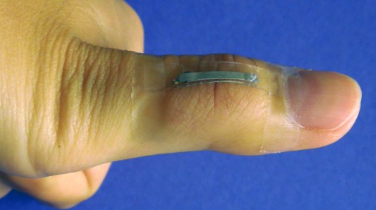
North Carolina State University researchers have developed a thumb joint mounted, multifunctional sensor using silver nanowires that measures strain, pressure, human touch and bioelectronic signals.  With potential biomedical, military and athletic applications, the sensor can be used for prosthetics, robotic systems and flexible touch panels.

“The technology is based on either physical deformation or “fringing” electric field changes. The latter is very similar to the mechanism used in smartphone touch screens, but the sensors we’ve developed are stretchable and can be mounted on a variety of curvilinear surfaces such as human skin” according to NC State’s Shanshan Yao.

“These sensors could be used to help develop prosthetics that respond to a user’s movement and provide feedback when in use,” said Professor Yong Zhu. “They could also be used to create robotics that can ‘feel’ their environment, or the sensors could be incorporated into clothing to track motion or monitor an individual’s physical health.”

Categories
Diabetes mHealth Monitoring Sensors

Smart contact lens prototype to monitor glucose

http://googleblog.blogspot.com/2014/01/introducing-our-smart-contact-lens.html

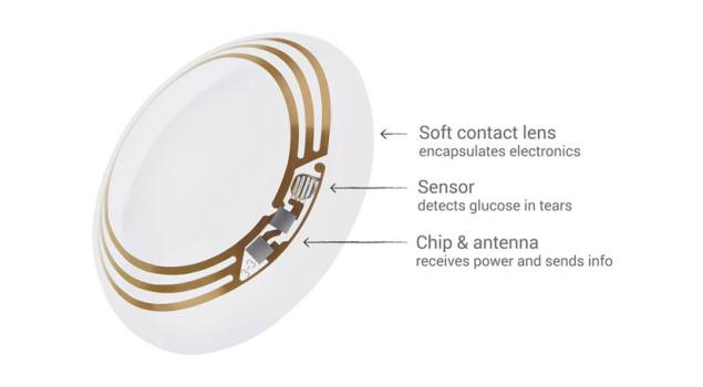
Google is testing a smart contact lens that’s built to measure glucose levels in tears.  It uses a tiny wireless chip and miniaturized glucose sensor embedded between two layers of soft contact lens material. The prototype can generate a reading once per second. They are investigating the potential of integrating tiny LED lights that could light up to indicate that glucose levels have crossed above or below thresholds.  Google says it is working with the FDA to turn the prototypes into products.

Categories
AI BCI Brain

K supercomputer runs largest neural simulation to date

http://www.telegraph.co.uk/technology/10567942/Supercomputer-models-one-second-of-human-brain-activity.html

RIKEN, Okinawa Institute of Science and Technology, and Forschungszentrum Jülich researchers have used Japan’s K computer to run the longest brain simulation to date.With 705,024 processor cores, running at speeds of over 10 petaflops, it is ranked the world’s fourth-most powerful computer. Using Neural Simulation Technology software and 92,944 of its processors, the computer replicated one second of brain activity across 1.73 billion nerve cells and 10.4 trillion synapses.  This represents one per cent of the brain’s neuronal network and took the K computer 40 minutes.

“The new result paves the way for combined simulations of the brain and the musculoskeletal system using the K computer,” said Kenji Doya of the Okinawa Institute of Science of Technology,. “These results demonstrate that neuroscience can make full use of the existing peta-scale supercomputers.”

“If peta-scale computers like the K computer are capable of representing one per cent of the network of a human brain today, then we know that simulating the whole brain at the level of the individual nerve cell and its synapses will be possible with exa-scale computers hopefully available within the next decade,” said project leader Markus Diesmann.

Categories
Heart

Study: Surgeons view detailed, real-time 3D holograms of heart

http://www.realviewimaging.com/

Philips and RealView Imaging recently completed a clinical study demonstrating the feasibility of using live 3D holographic visualization and interaction technology to guide minimally-invasive structural heart disease procedures. RealView’s technology was used to display interactive, real-time 3D holographic images acquired by Philips’ interventional X-ray and cardiac ultrasound systems.

In addition to viewing the patient’s heart on a 2D screen, doctors were able to view detailed dynamic 3D holographic images of the heart ’floating in free space’ during a minimally-invasive structural heart disease procedure, without using special eyewear. They were able to manipulate the projected 3D heart structures by touching the holographic volumes in front of them. The study demonstrated the potential of the technology to enhance the context and guidance of structural heart repairs.

The Yokneam, Israel based company says it plans to launch the system in 2015.

Categories
Brain EEG Ultrasound

Ultrasound stimulation enhanced sensory performance in human brain

http://www.nature.com/neuro/journal/vaop/ncurrent/full/nn.3620.html

Virginia Tech Carilion Research Institute scientists, led by Professor William Tyler, have demonstrated that ultrasound directed to a specific region of the brain can boost performance in sensory discrimination. This is the first example of low-intensity, transcranial-focused ultrasound modulating human brain activity to enhance perception.

The scientists delivered focused ultrasound to an area of the cerebral cortex that corresponds to processing sensory information received from the hand. To stimulate the median nerve, they placed an electrode on the wrist and recorded brain responses using EEG. Before stimulating the nerve, they began delivering ultrasound to the targeted brain region.  The ultrasound decreased the EEG signal and weakened the brain waves responsible for encoding tactile stimulation.

Subjects were then given two neurological tests: the two-point discrimination test, which measures a one’s ability to distinguish whether two nearby objects touching the skin are truly two distinct points, rather than one; and the frequency discrimination task, which measures sensitivity to the frequency of a chain of air puffs.  The subjects receiving ultrasound showed significant improvements in their ability to distinguish pins at closer distances and to discriminate small frequency differences between successive air puffs.

Categories
Eyes Sensors Wearables

Smart contact lens with potential to monitor intraocular pressure

http://www.nature.com/ncomms/2014/140107/ncomms3982/full/ncomms3982.html

Swiss Federal Institute of Technology scientists have developed a light, flexible, ultra-thin membrane with the potential to detect intraocular pressure in glaucoma.  Researchers claim that the technology “could offer significant advantages over existing solutions in terms of thickness, lightness, and transparency and, hence, comfort for the patient.”

The device consists of layered polymer films, one of which is a semiconductor. Gauge sensors monitor intraocular pressure in response to strain, which detects the high eye pressure typical of glaucoma.  In trials, the scientists transferred the material onto plastic contact lenses on an artificial eye.

The researchers must overcome technical obstacles before their solution is commercially viable. The method of attaching electronics to the contact lens must be optimized to include effects of the aqueous ocular environment.  Additionally, in the lab, the required energy is provided from an external source.  A different solution would be needed for a unit attached to a human eye.

Categories
Sensors Sleep

Nerve stimulation for sleep apnea

http://www.nejm.org/doi/full/10.1056/NEJMoa1308659

Current sleep apnea treatments are intrusive and uncomfortable. University of Pittsburgh Professor Patrick J. Strollo has developed a  “pulse generator” which, when surgically implanted, senses one’s effort to breath.  It then sends pulse stimulation to the hypoglossal nerve, controlling the neck muscles that keep the airway open, preventing collapse.

Standard treatment for sleep apnea is a device called a CPAP. It is a machine that applies air pressure through a mask, which, if worn while sleeping, keeps the airway open. Many people find the masks bulky and uncomfortable, choosing instead to leave their apnea untreated.