Categories
Brain

Precision PET scanner corrects patient movements, improving accuracy

http://www.hamamatsu.com/jp/en/news/development/20130905000001.html

Japan’s Hamamatsu Photonics and Hamamatsu University School of Medicine are developing a new PET diagnostic system for Alzheimer’s disease and other brain disorders.  The system combines a tracer that reflects changes in acetylcholine-related cognitive functions and a high-precision brain PET scanner that can correct the movement of a patient’s head.  It is designed to improve the accuracy and overcome the limitations of current systems, which require patients to not move for a long period of time.

Categories
Brain EEG

Biofeedback tool identifies seizure brain patterns through music

http://news.stanford.edu/news/2013/september/seizure-music-research-092413.html

In a recent experiment, Stanford professors Chris Chafe and Josef Parvizi created audio EEG recordings of both normal brain activity and seizure states.  During the state of seizure, tones became more pronounced and their tempo became chaotic.  “We could instantly differentiate seizure activity from non-seizure states with just our ears,” Chafe said. “It was like turning a radio dial from a static-filled station to a clear one.”

Since some seizures can occur without immediate, behavioral symptoms, Chafe and Parvizi have decided to use this research to develop a tool for caregivers to use real time brain data to hear and recognize undetected seizures.

The EEGs Parvizi conducts register brain activity from more than 100 electrodes.  Chafe selects certain electrode/neuron pairings and allows them to modulate notes sung by a female singer. As the electrode captures increased activity, it changes the pitch and inflection of the singer’s voice.

Before the seizure begins (during the pre-ictal stage) the notes from each “singer” almost synchronize and fall into a clear rhythm.  In the moments leading up to the seizure event, each of the singers begins to improvise. The notes become progressively louder and more scattered as the full seizure event occurs (the ictal state).  One can hear the electrical storm originate on one side of the brain and eventually cross over into the other hemisphere.  After about 30 seconds of chaos, the singers begin to calm, tapering off into their post-ictal rhythm. Occasionally, one or two will behave erratically, but on the whole, the choir sounds extremely fatigued.

According to Professor Parvizi, this is the perfect representation of the three phases of a seizure event.

Categories
Brain

High speed fluorescent camera for blood diagnostics, brain mapping

http://www.nature.com/nphoton/journal/v7/n10/full/nphoton.2013.245.html

UCLA Professor Bahram Jalali has developed a high-speed microscopy technique, forming images by reading an entire row of pixels at once and encoding the fluorescence from each pixel on a different radio frequency. The camera forms images approximately 10 times faster than current state-of-the-art technologies.
A laser beam is first split into two beams, with one of the beams changed slightly in frequency. The two beams then combine again on the sample that has been labeled with fluorescent molecules. As they interfere at the sample, the frequency difference between the two beams encodes the fluorescence from each pixel, allowing the camera can detect an entire row of pixels at one time. The researchers call the new technique “fluorescence imaging using radiofrequency-tagged emission,” or FIRE.
The technique can be applied in flow cytometry, which analyzes cells one by one in a flowing fluid. Unlike current imaging flow cytometers, the FIRE technique can keep up with conventional flow-cytometry speeds and image 50,000 cells per second. While most flow cytometers take only a single-point estimate of a cell’s fluorescence, FIRE can create an entire high-resolution, multicolor image of each cell, providing significantly more information to a biologist or clinician.
Lead author Eric Diebold said that “this information could be the difference between detecting a rare cancer cell in a blood sample or missing it completely”  and that they can “capture images of cells that are blur-free as they flow by at a speed around 100 times faster than state-of-the-art imaging flow cytometers.”
The researchers claim that the technique can also be applied in vivo, including viewing neural activity in the brain, making a much larger field of view available for analysis.
Categories
Assistive Technologies BCI Brain Sensors

Mind controlled bionic leg

http://www.nejm.org/doi/full/10.1056/NEJMoa1300126

A robotic control system for a prosthetic leg allowed a 31-year-old man to walk and climb stairs with a nearly normal gait. The system links nerves in the thigh — including some for missing muscles in the lower limb — to a processor that decodes the signals and guides the motion of the prosthesis, according to Levi Hargrove of the Rehabilitation Institute of Chicago.

The prosthetic limb includes thirteen mechanical sensors and can be used — like many commercially available prostheses — by changing its settings with a wireless key fob. Combining electromyographic signals from the residual limb and the sensor data eliminated the need to change settings with an external device and produced unique stride patterns for each type of ambulation, such as walking on a ramp and climbing stairs.  Adding the data from the nerves to the information from the sensors reduced the error rate — misclassification by the control system of the patient’s intended movement — from 12.9% to 1.8% of all motions.

Categories
BCI Brain fMRI

Neurofeedback enhances signal-to-noise ratio in thought

http://research.vtc.vt.edu/news/2013/sep/13/covert-operations-your-brain-digitally-remastered/.

Virgina Tech Carillon Professor Stephen Laconte developed technology to transfer non-invasive brain activity measurements into control signals that drive physical devices and computer displays in real time. The study suggests that the signal-to-noise ratio of the brain activity underlying our thoughts can be remastered. Researchers used whole-brain, classifier-based real-time fMRI to understand the neural underpinnings of brain-computer interface control.

24 subjects were asked to control a visual interface by silently counting numbers at fast and slow rates. For half the tasks, the subjects were told to use their thoughts to control the movement of the needle on the device they were observing; for the other tasks, they simply watched the needle.  Scientists discovered a feedback effect: the subjects who were in control of the needle achieved a better whole-brain signal-to-noise ratio than those who simply watched the needle move.  The act of controlling the computer-brain interface also led to an increased classification accuracy, which corresponded with improvements in the whole-brain signal-to-noise ratio.

This enhanced signal-to-noise ratio has implications for brain rehabilitation.

Categories
Assistive Technologies BCI Brain Conference Seniors Wearables

Data glasses controlled by eye movement — an alternative to brain machine interface

http://www.domain-b.com/technology/20130912_movements.html
Researchers at the Fraunhofer Institute have developed bidirectional OLED microdisplay eye-controlled data glasses.  Users can view the real world while browsing a large amount of virtual information and turn pages with their eyes.

Integrated camera sensors register the direction of the wearer’s eye movements and an image processing program calculates the exact position of their pupils in real time.  An infrared light source in the glass frame produces accurate positioning results even in low light.

The glasses can enable elderly and disabled people to attract attention in emergencies using nothing but their eyes – or simply provide a way to change the TV channel using specific eye movements.

Categories
BCI Brain

Duke researchers link visual stimulus with tactile sensation

http://www.dukehealth.org/health_library/news/touch-and-movement-neurons-shape-the-brain-s-internal-image-of-the-body
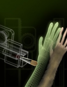
Miguel Nicolelis is one of the main contributors to brain machine interface.  In a series of innovative experiments, he demonstrates the intricate connections in the brain, attempting to create a coherent model of multi-sensory input.  His recent experiment shows that monkeys can be tricked when the multi-sensory input is only partially coherent.

A related study from Stockholm’s Karolinska Institute describes humans distorted self perception due to incoherency between visual and tactile input.

In the Nicolelis study, untrained monkeys were implanted with up to 384 electrodes to analyze how stimulus was encoded by the monkey brain. The monkeys were then shown a virtual simulation of their arm being touched, while simultaneously having their own arm touched. After a few minutes of synchronized stimulation, the physical component was removed. The areas of the monkeys’ brains responsible for tactile sensations, however, continued to respond to the virtual simulation, indicating that they felt physical sensation simply through visual association. The monkeys’ neuronal responses to the virtual stimulation came later than the responses to the physical stimulation. This suggests that the sensation was mediated by a longer pathway involving the visual system.

Instead of previously imagined single neuronal pathways, seemingly unrelated cortices apparently use a highly dynamic, cross functional process to form more of a continuously interacting grid or network. They also appear to cooperate quite closely in shaping the body schema, or the brain’s internal representation of the body

Categories
Brain Seniors

Video game improves cognition in seniors

http://www.ucsf.edu/news/2013/09/108616/training-older-brain-3-d-video-game-enhances-cognitive-control
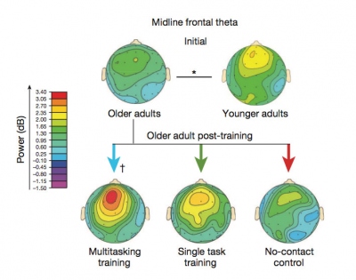
UCSF researchers found that older adults improved cognitive controls, including multitasking and the ability to sustain attention, by playing a specially designed videogame — and that the effects can be long lasting.

In the game, participants race a car around a winding track while a variety of road signs pop up. Drivers are instructed to keep an eye out for a specific type of sign, while ignoring all the rest, and to press a button whenever that particular sign appears.  The need to switch rapidly from driving to responding to the signs – i.e. multitasking – generates interference in the brain that undermines performance. The researchers found that this interference increases dramatically across the adult lifespan.

The study found that after training, the older adults were able to perform at a higher level than untrained 20-year-olds and that the positive effects lasted for at least six months.

Scientists continue to explore the connection between videogames and the brain’s information-processing functions, such as memory, attention, decision-making and creativity.

Categories
Brain

Imaging technology distinguishes between brain tissue and tumors at microscopic level

http://stm.sciencemag.org/content/5/201/201ra119
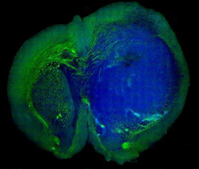
Harvard’s Xiaoliang Sunney Xie and Minbiao Ji used SRS microscopy (Stimulated Raman Scattering) to “see” the tiniest areas of tumor cells in brain tissue, and to distinguish tumor from healthy tissue in the brains of living mice.  They then showed that the same was possible in tissue removed from a patient with glioblastoma multiforme, one of the most deadly brain tumors.

Professor Xie described that “Biopsy has been the gold standard for detecting and removing these types of tumors.  But this technique, we believe, is better because it’s live. Surgeons can now skip all the steps of taking a biopsy, freezing, and staining the tissue. This technique allows them to do it all in vivo.”

SRS works by shining non-invasive lasers into tissue and detecting the weak signal that emerges. By analyzing the signal’s spectrum, researchers can build images of the cellular makeup of the tissue. By amplifying those signals, they transform a technique that once took hours or days into one that works in real time, and could offer a critical insight to surgeons in the operating room. Since brain tissue and tumors contain different chemical makeups, researchers can create images that precisely show where the tumor “margin” — the boundary area where tumor cells infiltrate among normal cells — is located, helping to guide surgeons in the operating room.

Categories
Brain

Vienna scientists create 3-D human brain tissue from stem cells

http://www.technologyreview.com/news/518716/researchers-grow-3-d-human-brain-tissues/

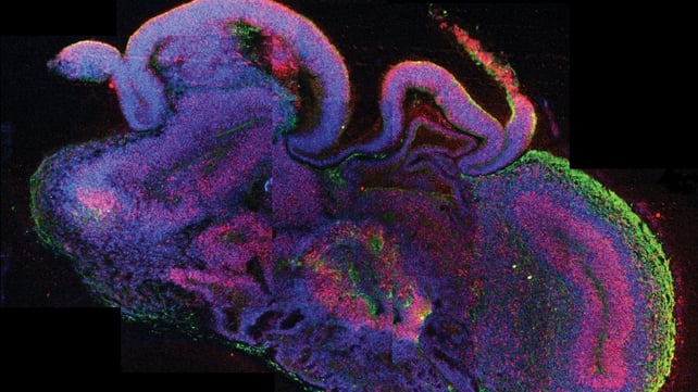
Scientists at the Austrian Academy of Sciences have turned human stem cells into pea-sized mini-brains with a neural structure similar to the brain of a developing embryo.  These “cerebral organoids”, as they are termed formally, are the best living model of a human brain created to date.

The researchers have already used their mini-brains to investigate one neuronal disorder, microcephaly, in which the brain does not grow properly. They hope to apply the technique to more complex conditions such as autism and schizophrenia, for which no good animal models are available.
Categories
BCI Brain Conference

Human-to-human brain interface – UW researcher controls colleague’s movement

http://www.washington.edu/news/2013/08/27/researcher-controls-colleagues-motions-in-1st-human-brain-to-brain-interface/

University of Washington researchers have performed what they believe is the first noninvasive human-to-human brain interface, with one researcher able to send a brain signal via the Internet to control the hand motions of a fellow researcher.

Using electrical brain recordings and a form of magnetic stimulation, Rajesh Rao sent a brain signal to Andrea Stocco on the other side of the UW campus, causing Stocco’s finger to move on a keyboard.

While researchers at Duke University have demonstrated brain-to-brain communication between two rats, and Harvard researchers have demonstrated it between a human and a rat, Rao and Stocco believe this is the first demonstration of human-to-human brain interfacing.

Magnetic stimulation as a direct brain communication channel is very intriguing.

Categories
AI Apps Assistive Technologies Brain mHealth Monitoring Seniors Sensors

Personalized robot companion for seniors

http://cordis.europa.eu/fetch?CALLER=OFFR_TM_EN&ACTION=D&RCN=11525

A European consortium of research institutes, universities and technology companies has developed a highly customizable robot companion to help seniors to maintain their quality of life, stay healthy and avoid social exclusion.

The robot, a mobile wheeled semi-humanoid figure equipped with cameras, sensors, audio, and a touch screen interface, can remind users to take their medicine, suggest they have their favorite drink, or prompt them to go for a walk or visit friends if they haven’t been out for a while. As part of a larger smart-home environment that can include smart clothing to monitor vital signs, the system can monitor user’s health and safety, and alert emergency services if necessary.