Categories
Autism Brain MRI

Prenatal MRI study suggests autism differences may begin in the womb

A small Boston Children’s Hospital study led by Assistant Professor Emi Takahashi and postdoc Alpen Ortug showed increased volume of the insular lobe as a potential strong prenatal MRI biomarker that could predict the emergence of ASD later in life. It revealed significant differences in brain structures at 25 weeks’ gestation between children who were later diagnosed with ASD and those who were not.

If validated, this could enable earlier treatment after birth, which could improve outcomes. Early treatment has been shown to improve language and cognitive abilities, but current diagnostic tools can only identify the disorder around 18 months of age.

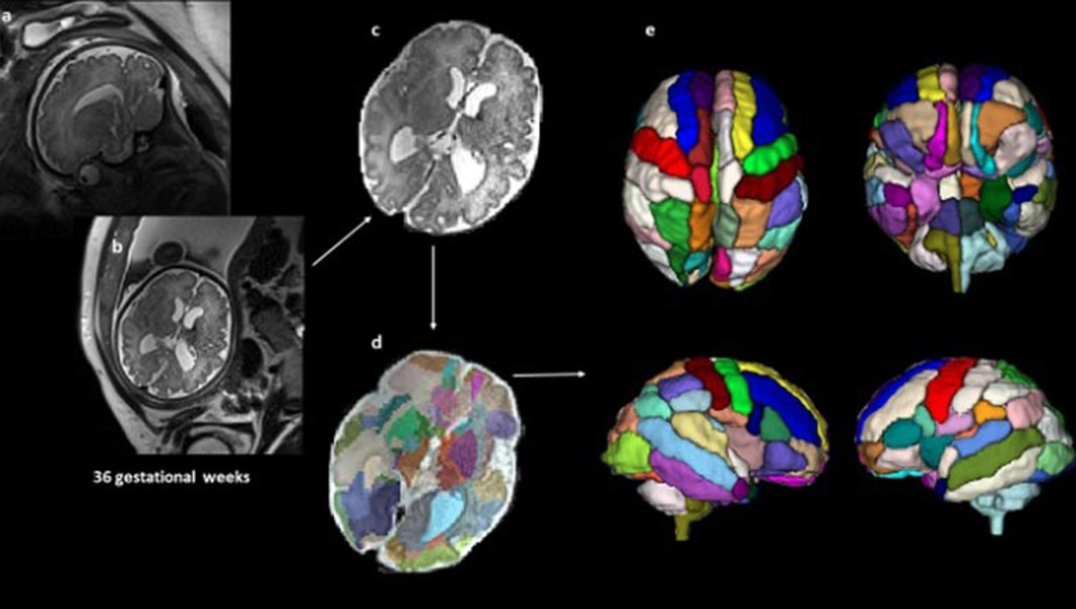
39 fetal MRI brain scans at 25 weeks’ gestation taken at Boston Children’s Hospital were analyzed. 9 children were later diagnosed with ASD, 20 were neurotypical and 10 did not have ASD but had other conditions also observed in the children with ASD.

An atlas-based automated anatomical labeling method was used to segment the scans and compare brain regions between the different groups. The insular lobe was found to have significantly larger volume in the ASD group compared with the other 3 control groups.

The findings align with studies that have reported changes in the insular cortex in adults with autism, and suggests these differences may begin in the womb. The scans from children with ASD also showed a significantly larger amygdala and hippocampal commissure compared with children who had other health conditions but not ASD.

According to Ortug, “to the best of our knowledge, this is the first attempt to semi-automatically segment the brain regions in the prenatal stage in patients who are diagnosed with autism later and compare different groups of controls.”


Join ApplySci at MIT on September 30th for Deep Tech Health + Neurotech Boston 2022
Categories
AI MRI

AI speeds MRI scans

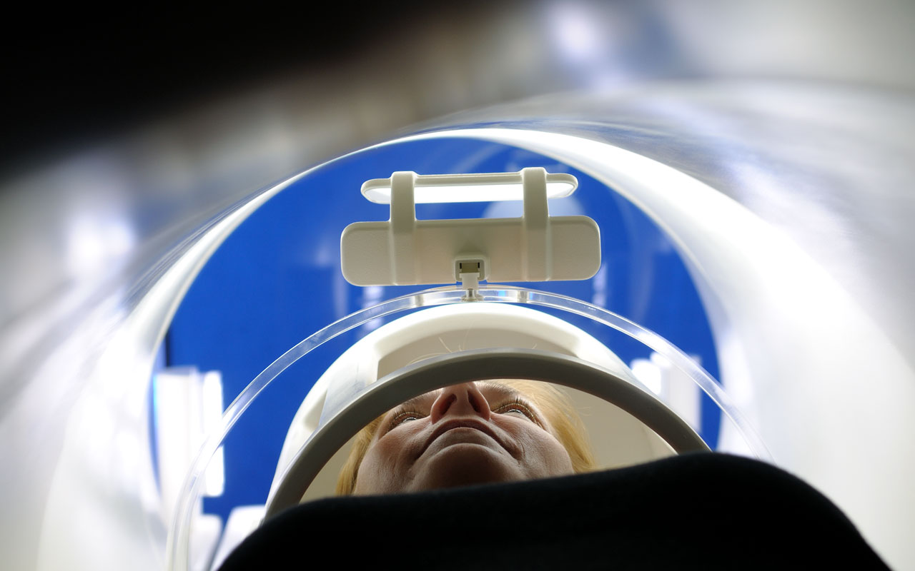
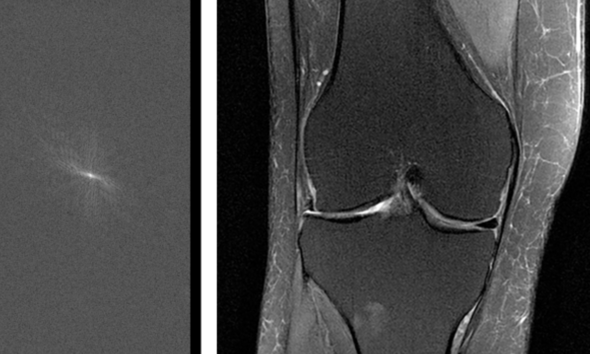
Facebook and NYU’s fastMRI project, led by Larry Zitnick, uses AI in an attempt to make MRI imaging 10 times faster. Neural networks will be trained to fill in missing or degraded parts of scans, turning them from low resolution into high. The goal is to significantly reduce the time patients must lie motionless inside an MRI machine.


Join ApplySci at the 9th Wearable Tech + Digital Health + Neurotech Boston conference on September 24, 2018 at the MIT Media Lab.  Speakers include:  Rudy Tanzi – Mary Lou Jepsen – George ChurchRoz PicardNathan IntratorKeith JohnsonJuan EnriquezJohn MattisonRoozbeh GhaffariPoppy Crum – Phillip Alvelda Marom Bikson – Ed Simcox – Sean Lane

Categories
Conference MRI

Ultra-low-field, portable MRI

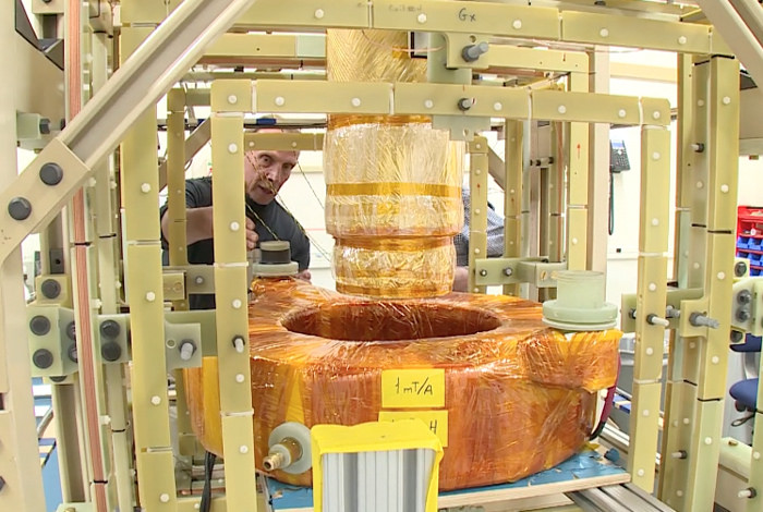
Los Alamos National Laboratory‘s Michelle Espy is developing an ultra-low-field, lightweight MRI system for use on the battlefield and in poor countries.  The device will be simple to transport, set up, and use in non-traditional settings.

Conventional MRI machines use large magnetic fields that align protons in water molecules. Magnetic resonance signals are detected and turned into images. Highly detailed images are created, but the process is complicated and expensive. Espy uses Superconducting Quantum Interference Devices (SQUID) to create quality images with ultra-low-magnetic fields.

The  first generation (battlefield) “b”MRI was built in a large metal housing to shield it from interference.  The team is now surrounding the system with lightweight wire coils in the open environment to compensate the Earth’s magnetic field.  A field compensation system will soon eradicate invading magnetic field signals.

WEARABLE TECH + DIGITAL HEALTH NYC 2015 – JUNE 30 @ NEW YORK ACADEMY OF SCIENCES. REGISTER HERE.

Categories
MRI Seniors

High speed MRI analyzes vocal movement

Aaron Johnson of The Beckman Institute has developed an MRI technique that can view dynamic images of vocal movement at 100 frames per second.  This speed is far more advanced than any other MRI technique.  The method is especially useful in studying how rapidly the tongue moves, along with other muscles in the head and neck, during speech and singing.  The attached video demonstrates the results.

To combine the imaging with audio, the researchers used a noise-canceling fiber-optic microphone to pull out the voice, and aligned the audio track with the imaging.

According to Johnson, the neuromuscular system and larynx change and atrophy with age, contributing to deficits associated with the older voice, such as a weak, strained, or breathy voice.  He wants to understand how these changes occur, and if interventions, such as vocal training, can reverse the effects. This requires seeing how the muscles of the larynx move in real time.

Wearable Tech + Digital Health NYC 2015 – June 30 @ New York Academy of Sciences.  Early registration discount ends Friday, 4/24.