Categories
Autism Brain MRI

Prenatal MRI study suggests autism differences may begin in the womb

A small Boston Children’s Hospital study led by Assistant Professor Emi Takahashi and postdoc Alpen Ortug showed increased volume of the insular lobe as a potential strong prenatal MRI biomarker that could predict the emergence of ASD later in life. It revealed significant differences in brain structures at 25 weeks’ gestation between children who were later diagnosed with ASD and those who were not.

If validated, this could enable earlier treatment after birth, which could improve outcomes. Early treatment has been shown to improve language and cognitive abilities, but current diagnostic tools can only identify the disorder around 18 months of age.

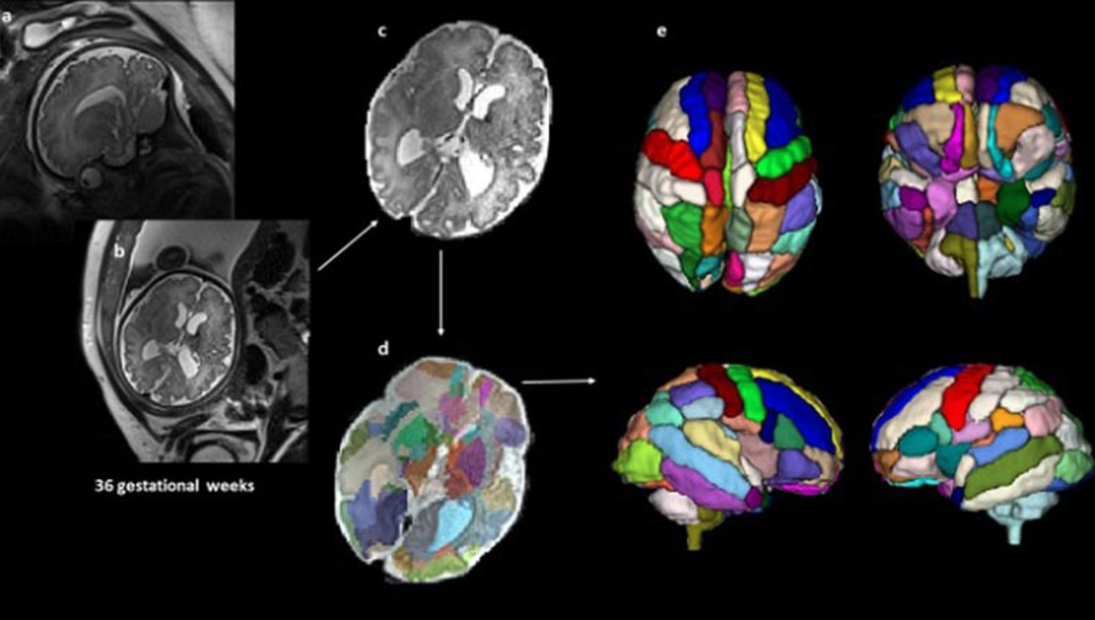
39 fetal MRI brain scans at 25 weeks’ gestation taken at Boston Children’s Hospital were analyzed. 9 children were later diagnosed with ASD, 20 were neurotypical and 10 did not have ASD but had other conditions also observed in the children with ASD.

An atlas-based automated anatomical labeling method was used to segment the scans and compare brain regions between the different groups. The insular lobe was found to have significantly larger volume in the ASD group compared with the other 3 control groups.

The findings align with studies that have reported changes in the insular cortex in adults with autism, and suggests these differences may begin in the womb. The scans from children with ASD also showed a significantly larger amygdala and hippocampal commissure compared with children who had other health conditions but not ASD.

According to Ortug, “to the best of our knowledge, this is the first attempt to semi-automatically segment the brain regions in the prenatal stage in patients who are diagnosed with autism later and compare different groups of controls.”


Join ApplySci at MIT on September 30th for Deep Tech Health + Neurotech Boston 2022
Categories
Brain

Minimally invasive sensor detects electrical activity, optical signals in brain for MRI

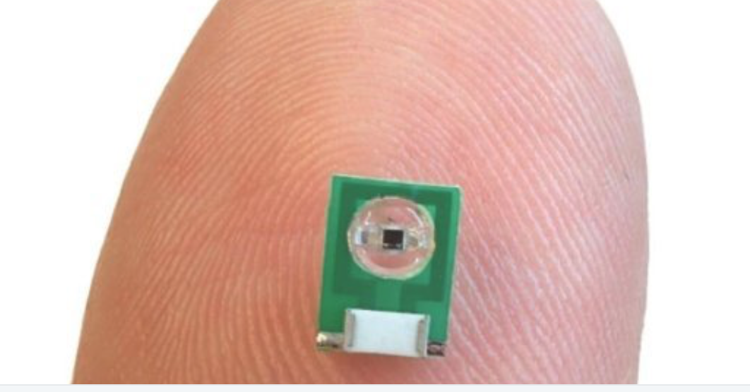
MIT’s Aviad Hai has developed a minimally invasive sensor to detect electrical activity or optical signals in the brain for MRI. No power source is needed, as radio signals that an external MRI scanner emits power the sensor.  It is implanted but does not require a wired connection to the brain. The researchers believe that it could also be adapted to measure glucose or other chemicals.

The team previously developed MRI sensors to detect calcium, serotonin and dopamine. The new sensor is meant to replace current electrical activity monitoring, which is extremely invasive, and can cause tissue damage.

Hai and colleagues shrank a radio antenna down to a few millimeters, so that it could be implanted directly into the brain to receive radio waves generated by water in the tissue.

The sensor is first tuned to the same frequency as the radio waves emitted by the hydrogen atoms. When an electromagnetic signal is detected, its tuning changes and  it no longer matches the hydrogen atom frequency. A weaker image then arises when the sensor is scanned by an external MRI machine.

In a study, the sensors were able to pick up electrical signals similar to those produced by action potentials or local field potentials.

Hai plans to further miniaturize the sensor, to enable multiple injections, to image light or electrical fields over a larger brain area.

Dr. Hai will discuss this work at ApplySci’s Wearable Tech + Digital Health + Neurotech Silicon Valley conference on February 21-22 at Stanford University


Join ApplySci at the 10th Wearable Tech + Digital Health + Neurotech Silicon Valley conference on February 21-22 at Stanford University — Featuring:  Zhenan BaoChristof KochVinod Khosla – Nathan IntratorJohn MattisonDavid EaglemanUnity Stoakes Shahin Farshchi – Emmanuel Mignot – Michael Snyder – Joe Wang – Josh Duyan – Aviad Hai

Categories
AI MRI

AI speeds MRI scans

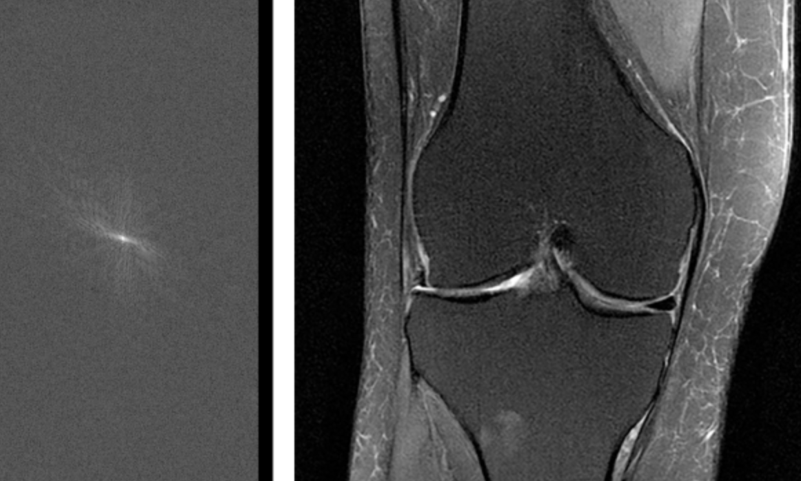
Facebook and NYU’s fastMRI project, led by Larry Zitnick, uses AI in an attempt to make MRI imaging 10 times faster. Neural networks will be trained to fill in missing or degraded parts of scans, turning them from low resolution into high. The goal is to significantly reduce the time patients must lie motionless inside an MRI machine.


Join ApplySci at the 9th Wearable Tech + Digital Health + Neurotech Boston conference on September 24, 2018 at the MIT Media Lab.  Speakers include:  Rudy Tanzi – Mary Lou Jepsen – George ChurchRoz PicardNathan IntratorKeith JohnsonJuan EnriquezJohn MattisonRoozbeh GhaffariPoppy Crum – Phillip Alvelda Marom Bikson – Ed Simcox – Sean Lane