Categories
Brain

Alzheimer’s diagnosis disputed in up to 50% of PET study subjects

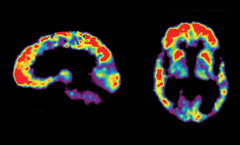
James Hendrix and Alzheimer’s Association colleagues are conducting a study to see how PET scans could change the nature of Alzheimer’s diagnosis and treatment.

4,000 of 18,000 subjects have been tested to date, with a stunning result showing that a significant portion of people with mild cognitive impairment or dementia who are taking medication for Alzheimer’s may not actually have the disease.

The PET scan determines if  MCI or dementia  patient brains contain the amyloid plaques that are one of the two hallmarks of Alzheimer’s disease. Among the 4,000 people tested, 54.3 percent of MCI patients and 70.5 percent of dementia patients had the plaques. A positive test for amyloid does not mean someone has Alzheimer’s disease, though their presence is thought to precede the disease and increase the risk of progression. A negative test means a person does not have the disease.

After seeing the PET imaging results, doctors changed their care plans for two-thirds of the patients in the study. According to Hendrix, “We thought we would be able to see about a 30 percent change, but we’re getting a 66 percent change, so it’s huge. We see high percentages of people who are on a drug and didn’t need to be on those drugs.”


Join ApplySci at Wearable Tech + Digital Health + NeuroTech Boston on September 19, 2017 at the MIT Media Lab – featuring  Joi Ito – Ed Boyden – Roz Picard – George Church – Nathan Intrator –  Tom Insel – John Rogers – Jamshid Ghajar – Phillip Alvelda – Michael Weintraub – Nancy Brown – Steve Kraus – Bill Geary – Mary Lou Jepsen – Daniela Rus

Registration rates increase Friday, July 28th.


ANNOUNCING WEARABLE TECH + DIGITAL HEALTH + NEUROTECH SILICON VALLEY – FEBRUARY 26 -27, 2018 @ STANFORD UNIVERSITY
 

Categories
Brain

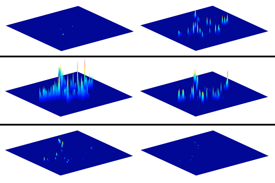
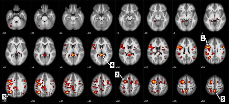
AI used to study brain blood flow ties to Schizophrenia

Mina Gheiratmand,  Serdar Dursun, and University of Alberta colleagues used IBM AI tools to try to  identify schizophrenic traits based on a person’s brain blood flow.

95 participant  fMRI images of schizophrenia-diagnosed and healthy patient brains were analyzed.  The researchers claimed to accurately diagnose patients, based on blood flow, 74% of the time. They also claimed to be able to measure the severity of symptoms.

A much larger sample size will be required to determine the validity of this approach to understanding a disease that so little is known about.  ApplySci applauds this perhaps promising attempt.

Categories
BCI Brain Ears Eyes

Direct brain path for sight, sound via implanted microscope

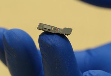
Rice University’s Jacob Robinson, with Yale and Columbia colleagues, are developing FlatScope — a flat, brain implanted microscope that monitors and triggers neurons which are modified to be fluorescent when active.

While capturing greater detail than current brain probes, the microscope also goes through deep levels that illustrate  sensory input processing — which they hope to be able to control.

Aiming to produce a super high-resolution neural interface, FlatScope is a part of  DARPA’s NESD program, founded by Phillip Alvelda, and now led by Brad Ringeisen.


Phillip Alvelda will be a featured speaker at ApplySci’s Wearable Tech + Digital Health + NeuroTech Boston conference on September 19, 2017 at the MIT Media Lab.  Other speakers include:  Joi Ito – Ed Boyden – Roz Picard – George Church – Nathan Intrator –  Tom Insel – John Rogers – Jamshid Ghajar  – Michael Weintraub – Nancy Brown – Steve Kraus – Bill Geary – Mary Lou Jepsen – Daniela Rus

Registration rates increase Friday, July 21st

Categories
Babies Brain EEG Pain

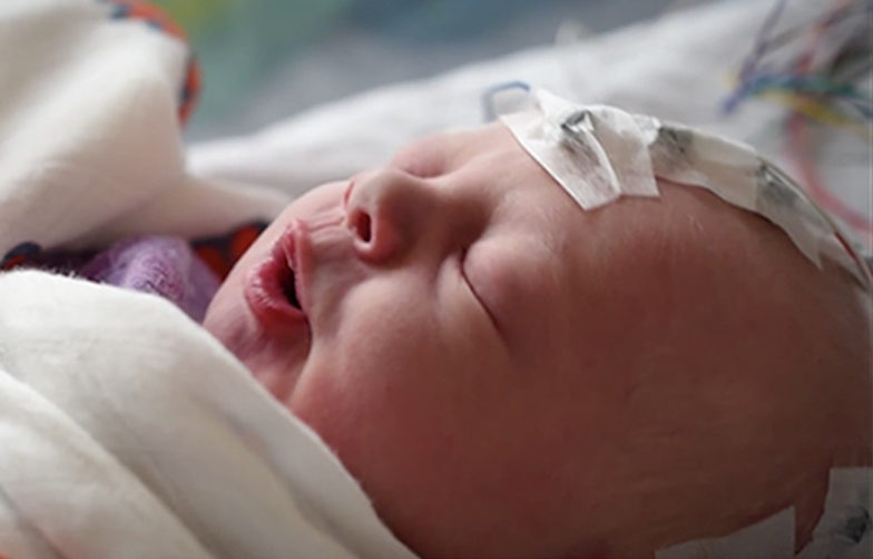
EEG detects infant pain

Caroline Hartley and Oxford colleagues studied 72 infants during painful medical procedures.  Using EEG, they found a signature change in brain activity about a half-second after a painful stimulus. They seek to understand its use in monitoring and managing infant pain, as well as  the use of EEG in adult pain treatment.

EEG is more precise than current heart rate, oxygen saturation level, and facial expression pain assessment, which are affected by other stressful, non-painful events.

In one experiment, 11 out of 12 infants had a decreased pain-related EEG signal after doctors applied a topical anesthetic to their feet.  A new study uses EEG to test the efficacy of morphine in infants, whose skin and intestines absorb drugs differently than adults.

EEG is being miniaturized by companies such as Neurosteer, making it an increasingly viable option for continuous pain, attention, and consciousness monitoring and treatment optimization.


Join ApplySci at Wearable Tech + Digital Health + NeuroTech Boston on September 19, 2017 at the MIT Media Lab – featuring  Joi Ito – Ed Boyden – Roz Picard – George Church – Nathan Intrator –  Tom Insel – John Rogers – Jamshid Ghajar – Phillip Alvelda – Michael Weintraub – Nancy Brown – Steve Kraus – Bill Geary


Categories
Brain Virtual Reality

VR studied for PTSD, phobia treatment

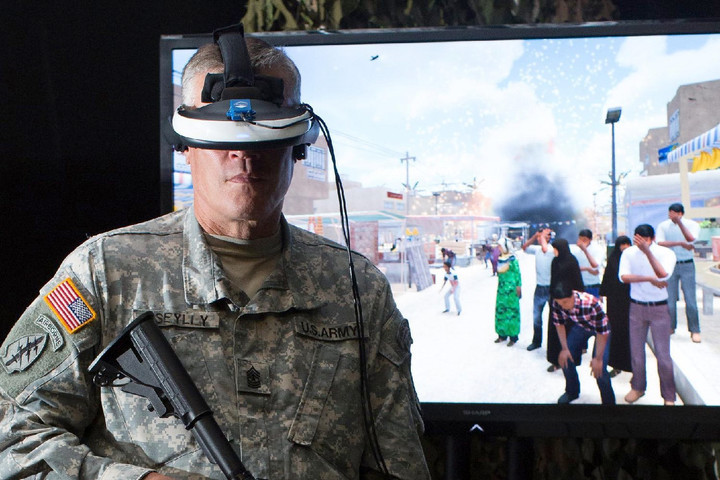
Emory’s Jessica Maples-Keller has published a study demonstrating the effectiveness of VR in treating PTSD, phobias, and other mental illnesses.  She describes the treatment as allowing “providers to create computer-generated environments in a controlled setting, which can be used to create a sense of presence and immersion in the feared environment for individuals suffering from anxiety disorders.”

Small studies on the use of VR in  panic disorder, schizophrenia, acute and chronic pain, addiction, and eating disorders have been done, but with limited numbers and a lack of comparison groups. Keller noted that extensive training is needed before integrating VR approaches into clinical practice.


Join ApplySci at Wearable Tech + Digital Health + NeuroTech Boston on September 19, 2017 at the MIT Media Lab. Featuring Joi Ito – Ed Boyden – Roz Picard – George Church – Nathan Intrator –  Tom Insel – John Rogers – Jamshid Ghajar – Phillip Alvelda

REGISTER BY MAY 19TH AND SAVE $500

Categories
Brain Heart Seniors Sensors

Wall sensor monitors walking speed, stride to track health

MIT’s Dina KatabiChen-Yu Hsu, and colleagues have developed WiGait, a wall sensor that detects walking speed and stride to monitor health. This builds on previous MIT research which showed that radio signals could track breathing and heart rate, without wearables.

The  system works by transmitting low-power radio signals and analyzing how they reflect off  bodies within a radius of 9 to 12 meters. Machine learning algorithms separated walking periods from other activities and found the stable phase within each walking period.  The sensor, when combined with wearable devices, could also track Parkinson’s and MS symptoms, and help predict health events related to  heart failure,  lung disease, kidney failure, and stroke, as well as the risk of falls. Caregivers could also be notified in emergencies.


Join ApplySci at Wearable Tech + Digital Health + NeuroTech Boston on September 19, 2017 at the MIT Media Lab. Featuring Joi Ito – Ed Boyden – Roz Picard – George Church – Tom Insel – John Rogers – Jamshid Ghajar – Phillip Alvelda – Nathan Intrator

Categories
Brain Sensors

Soft, flexible “skin-like” electrodes could improve brain interfaces

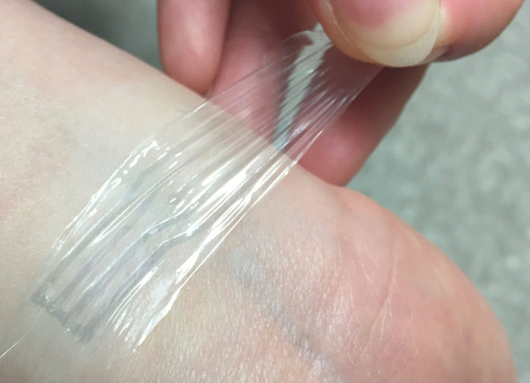
Stanford professor Zhenan Bao‘s latest flexible electronic initiative is the development of a plastic electrode that stretches like rubber but carries electricity like wires. This could be improve implanted brain interfaces which require soft and highly sensitive materials.

In a recent paper, Bao’s team describes the chemical modification of  brittle plastic to make it highly bendable, while enhancing  electrical conductivity. A more seamless connection between stiff electronics and flexible organic electrodes in our bodies is achieved.

According to lead author Yue Wang, “One thing about the human brain that a lot of people don’t know is that it changes volume throughout the day, It swells and deswells.”  Current electronic implants can’t stretch and contract with the brain, making it difficult to maintain a good connection.

Click to view Stanford University video.


Professor Bao was the keynote speaker at ApplySci’s recent Wearable Tech + Digital Health + NeuroTech conference at Stanford.

Join ApplySci at Wearable Tech + Digital Health + NeuroTech Boston – Featuring Roz Picard, Tom Insel, John Rogers and Nathan Intrator – September 19, 2017 at the MIT Media Lab
Categories
Brain Parkinson's

Carbon electrode technique tracks dopamine in the brain

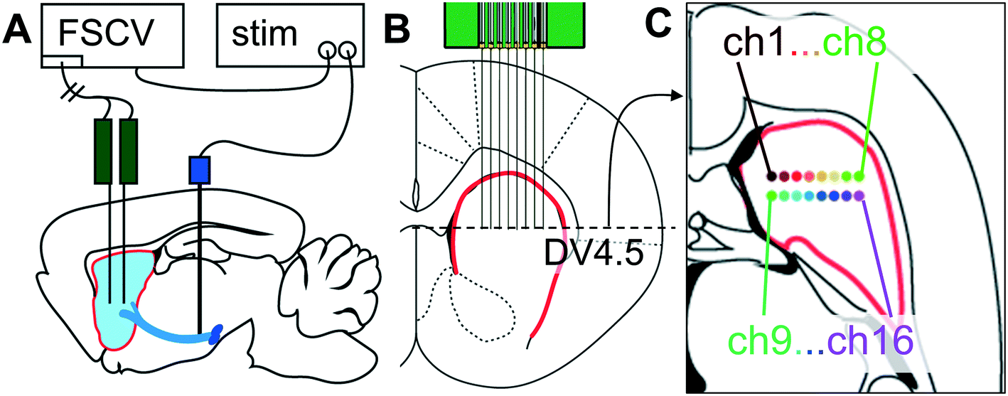
Michael Cima and MIT colleagues have developed a more precise tool to measure dopamine in the brain, to be able to study its role in in learning, memory, and emotion.

The new carbon electrode based technique can cover more of the brain, and provide longer, more accurate neurotransmitter readings, than previously possible.

The goal is a better understanding of neurtransmitter related diseases, and potential therapies to boost dopamine levels, in conditions that dysregulate it, such as Parkison’s disease.

According to lead author Helen Schwerdt: “Right now deep brain stimulation is being used to treat Parkinson’s disease, and we assume that that stimulation is somehow resupplying the brain with dopamine, but no one’s really measured that.”


Join ApplySci at Wearable Tech + Digital Health + NeuroTech Boston – Featuring Roz Picard, Tom Insel, John Rogers and Nathan Intrator – September 19, 2017 at the MIT Media Lab
Categories
BCI Brain

Magnetic coils might improve neural prostheses

Neocortex stimulation is used to treat neurological disorders, including Parkinson’s disease and paralysis. Current electrode-based implants have limited efficacy. It is difficult to create precise patterns of neural activity, or to achieve consistent responses over time.  This can be addressed by magnetic stimulation, but until now, coils small enough to be implanted into the cortex were not thought strong enough to activate neurons. Shelley Fried at Harvard has created a microcoil design that  has been effective for activating cortical neurons and driving behavioral responses.


Join ApplySci at Wearable Tech + Digital Health + NeuroTech Boston – Featuring Roz Picard, Tom Insel, John Rogers and Nathan Intrator – September 19, 2017 at the MIT Media Lab

Categories
Brain Robotics Stroke

Robots support neural and physical rehab in stroke, cerebral palsy

Georgia Tech’s  Ayanna Howard has developed Darwin, a socially interactive robot that encourages children to play an active role in physical therapy.

Specific targeting children with cerebral palsy (who are involved in current studies),  autism, or tbi, the robot is designed to function in the home, to supplement services provided by  clinicians.  It engages users as their human therapist would — monitoring performance, and providing motivation and feedback.In a recent experiment, motion trackers monitored user movements as Darwin offered encouragement, and demonstrated movements when they were not performed correctly.  Researchers claimed that wth the exception of one case, the robot’s impact was “significantly positive.

Darwin is still evolving (pun intended) and has not yet been commercialized.

At MIT,  Newman Lab researcher Hermano Igo Krebs has been using robots for gait and balance neurorehabilitation in stroke and cerebral palsy patients since 1989.  Krebs’s technology continues to be incorporated into  Burke Rehabilitation hospital treatment plans.


Join ApplySci at Wearable Tech + Digital Health + NeuroTech Boston – Featuring Roz Picard, Tom Insel, John Rogers and Nathan Intrator – September 19, 2017 at the MIT Media Lab
Categories
Autism Brain

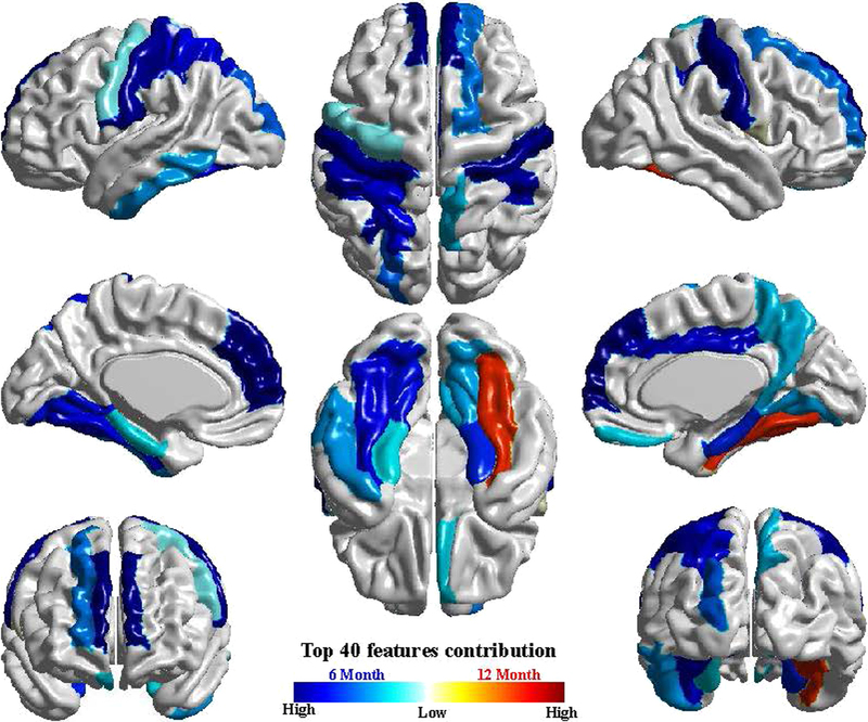
MRI, algorithm predict autism before behavioral symptoms appear

UNC’s Heather Hazlett has published a study showing that an overgrowth in brain volume, determined by MRI scans during the first year of life, forecasts whether a child at high risk of developing autism.

The goal is to give parents the opportunity to intervene long before behavioral symptoms become obvious, which usually occurs between ages 2 and 4.

The study was small, and further research is needed before it can be developed into a diagnostic  tool.

Two groups were studied: 106 high-risk  infants, with an older sibling with autism, and 42 low-risk infants, with no family history. MRI measurements of overall volume, surface area and thickness of the cerebral cortex in certain regions were done at set times between 6 and 24 months. An overgrowth of cortical surface area in infants later diagnosed with autism, compared with the typically developing infants, was discovered.

The researchers then developed an algorithm that predicted autism, based on brain measurements. Approximately 80% of the 15 high-risk infants who would later meet the criteria for autism at 24 months. Using the algorithm, the team also accurately predicted which babies would not develop autism by age 2.


Join ApplySci at Wearable Tech + Digital Health + NeuroTech Boston – Featuring Roz Picard, Tom Insel, John Rogers and Nathan Intrator – September 19, 2017 at the MIT Media Lab

Categories
Brain

Dopamine sensor tracks single neurons

MIT’s  Michael Strano has developed a carbon nanotube based detector that can track single cells’ secretion of dopamine. Using 20,000 sensor arrays, the team monitored dopamine secretion of single neurons, allowing them better understand dopamine dynamics.

Unlike most other neurotransmitters, dopamine can exert its effects beyond the synapse. Not all dopamine released into a synapse is taken up by the target cell, allowing some of the chemical to diffuse away and affect other nearby cells. Tracking this dopamine diffusion in the brain has proven difficult. Neuroscientists have tried using specialized electrodes, but only 20 of the smallest electrodes can be placed near any given cell.

The carbon nanotube sensors used in this study are coated with a DNA sequence that makes the sensors interact with dopamine. When dopamine binds to the carbon nanotubes, they fluoresce more brightly, allowing the researchers to see exactly where the dopamine was released. The researchers deposited more than 20,000 of these nanotubes on a glass slide, creating an array that detects any dopamine secreted by a cell placed on the slide.

According to Strano: “We have falsified the notion that dopamine should only be released at these regions that will eventually become the synapses,” Strano says. “This observation is counterintuitive, and it’s a new piece of information you can only obtain with a nanosensor array like this one.”


Join ApplySci at Digital Health + NeuroTech Boston – September 19, 2017 at the MIT Media Lab