
Categories
Brain Virtual Reality Wearables

VR + sensors improve accuracy, speed of PTSD diagnosis

PTSD is often misdiagnosed. Symptoms can be confused with those of depression.  Many clinicians lack the expertise needed to distinguish the condition, and therefore might not provide appropriate treatment.

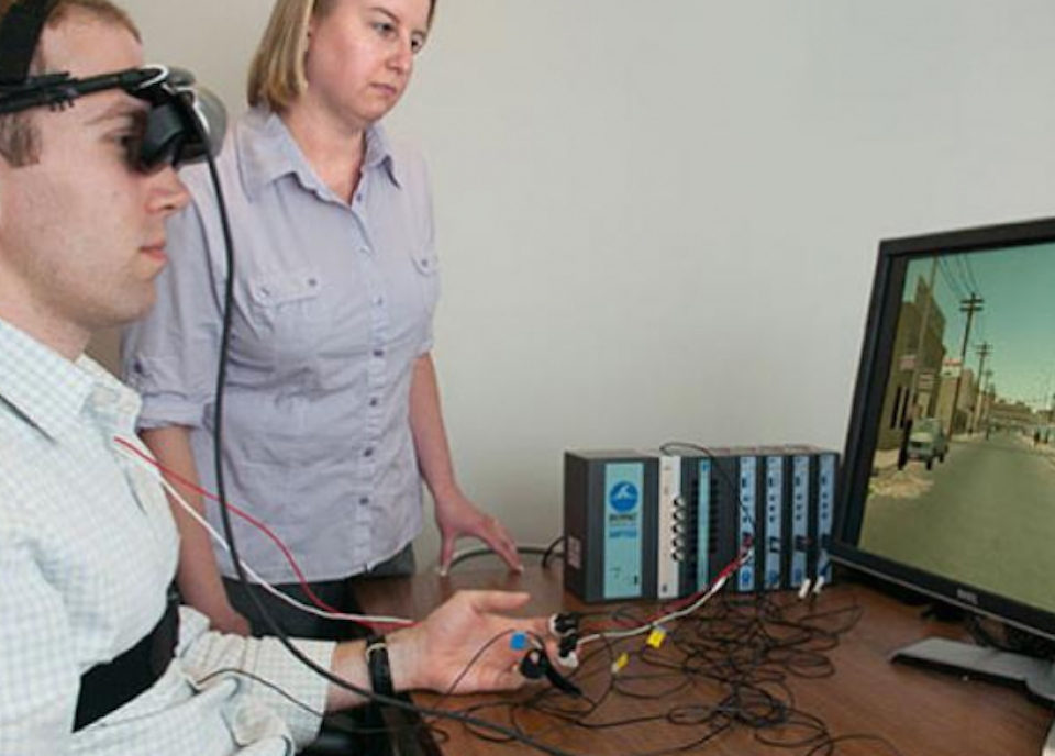
To address this widespread dilemma, Draper has developed a diagnostic system that combines virtual reality data with psychophysiological sensors. The sensors monitor heart rate, sweat, and pupil diameter, while subjects experience different types of audio and visual stimuli.

Stimuli customized to a patient’s personal traumatic experience can generate robust psychophysiological responses. However,  the time needed to tailor stimuli  is often not available in a point-of-care setting.  Draper’s solution uses generalized stimuli that results in quicker, more accurate assessments.

Additional research will address larger samples over a wide geographic area, as well as patients suffering from multiple mental health issue and  chronic diseases.

According to Dr. Philip Parks, who oversees Draper’s neurotechnology portfolio: “Once diagnosed with a particular disorder, such as depression, most mental health patients get relatively the same treatment even though their symptoms and response to treatment choices may be quite different. We hope that one day these technologies will help clinicians ensure that patients get the best possible medication and other treatments at the right time.”

Dr. Parks will be a featured speaker at NeuroTech NYC on June 8th at the New York Academy of Sciences.


Wearable Tech + Digital Health San Francisco – April 5, 2016 @ the Mission Bay Conference Center

NeuroTech San Francisco – April 6 @ the Mission Bay Conference Center

Wearable Tech + Digital Health NYC – June 7, 2016 @ the New York Academy of Sciences

NeuroTech NYC – June 8, 2016 @ the New York Academy of Sciences

Categories
BCI Brain

Brain state learning system adapts to user focus

BACh (Brain Automated Chorales) estimates brain workload using fNIRS to measure oxygen in the prefrontal cortex to help beginners learn to play Bach chorales.  The system offers new lessons when the brain isn’t overloaded with information.

Tufts Beste Yuksel and Robert Jacob, who developed the technology, believe that it can help with any type of learning, and specify math, engineering, programming, language and reading as examples.

In a recent study, 16 inexperienced piano players attempted to learn two chorales, one with the system’s assistance, and one with out. BACh first gave the musicians only the soprano line. When their cognitive load fell below a certain threshold, it added the bass part, then later the alto and tenor parts.  After 15 minutes, the pianists played more accurately and faster with BACh than without. Beginners saw more progress than intermediate level players.

The fNIRS machine is large, and Yuksel and Jacob are now working on a mobile system, which could incorporate emotion monitoring and feedback.


Wearable Tech + Digital Health San Francisco – April 5, 2016 @ the Mission Bay Conference Center

NeuroTech San Francisco – April 6, 2016 @ the Mission Bay Conference Center

Wearable Tech + Digital Health NYC – June 7, 2016 @ the New York Academy of Sciences

NeuroTech NYC – June 8, 2016 @ the New York Academy of Sciences
 

Categories
3-D Printing Brain

3D printed gel model replicates brain folding mechanism

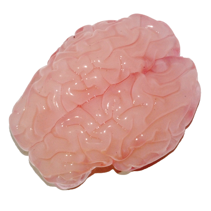
L. Mahadevan and Harvard colleagues have  used 3D printing to replicate a folding human brain.  The goal is to understand how brain folds are related to disease. While many molecular processes  determine cellular events, the study shows that what ultimately causes the brain to fold is a mechanical instability associated with buckling.
A 3D  gel model of a smooth fetal brain was created based on MRI images. To mimic cortical expansion, the gel brain was immersed in a solvent that is absorbed by the outer layer, causing it to swell relative to the deeper regions. The resulting compression led to the formation of folds similar in size and shape to real brains.
In humans, folding begins in fetal brains at the 20th week of gestation,  and is completed at a year and a half. The number, size, shape and position of neuronal cells during brain growth lead to the expansion of the cortex (gray matter), relative to the underlying white matter. The scientists said that this puts the cortex under compression, leading to a mechanical instability that causes it to crease locally. They believe that if a part of the brain does not grow properly, or if the global geometry is disrupted, the major folds may not be in the right place, which may cause dysfunction.
Click to view Harvard University GIF

Wearable Tech + Digital health San Francisco – April 5, 2016 @ the Mission Bay Conference Center

NeuroTech San Francisco – April 6, 2016 @ the Misson Bay Conference Center

Wearable Tech + Digital Health NYC – June 7, 2016 @ the New York Academy of Sciences

NeuroTech San Francisco – June 8, 2016 @ the New York Academy of Sciences

Categories
Apps Brain

Concussion app gauges recovery

As the incidence of and focus on concussion grows, the neurotech community continues to develop diagnostic tools and treatment protocols.

One such tool is the NYU developed Concussion Tracker app,  and corresponding study, designed to track self-reported physical and cognitive function after concussion.  The app does not, however, diagnose concussion.

The goal is to monitor recovery with greater detail and regularity,  to better evaluate treatment and identify persistent issues.   The project is led by NYU’s Laura BalcerDennis Cardone, and Paul Testa.

Testa said  the collected data will be used to  develop a new recovery evaluation app that in the future could shape recovery protocols.


Wearable Tech + Digital Health San Francisco – April 5, 2016 @ the Mission Bay Conference Center

NeuroTech San Francisco – April 6, 2016 @ the Mission Bay Conference Center

Wearable Tech + Digital Health NYC – June 7, 2016 @ the New York Academy of Sciences

NeuroTech NYC – June 8, 2016 @ the New York Academy of Sciences
 

Categories
BCI Brain

DARPA neural implant to enhance brain-computer connections

DARPA is leading the development of an improved  neural implant for connecting the brain to computers, using advances neuroscience, synthetic biology, low-power electronics, photonics and medical manufacturing.  Their goal is to to dramatically enhance  neurotechnology research capabilities and provide a foundation for new therapies.

The Neural Engineering System Design program aims to produce a miniaturized brain implant, smaller than one cubic centimeter in size, to improve data transfer. The  device would  translate between digital systems and the electrochemical “language” of the brain for more efficient communication.

NESD  is part of the BRAIN initiative and is led by Phillip Alvelda, who is “upgrading tools to really open the channel between the human brain and modern electronics.”

Current neural interfaces  use approximately 100 channels, each  aggregating signals from tens of thousands of neurons. The NESD program aims to develop technology to communicate directly with  one million individual neurons in a brain region.

Initial applications will include devices for those with sight or hearing impairments.  The system could feed digital auditory or visual information to the brain with  greater resolution and clarity than current technology.

Phillip Alveda will discuss this and other DARPA initiatives  at ApplySci’s NeuroTech San Francisco conference on April 6th.


Wearable Tech + Digital Health San Francisco – April 5, 2016 @ the Mission Bay Conference Center

NeuroTech San Francisco – April 6, 2016 @ the Mission Bay Conference Center

Wearable Tech + Digital Health NYC – June 7, 2016 @ the New York Academy of Sciences

NeuroTech NYC – June 8, 2016 @ the New York Academy of Sciences

 

Categories
Brain

Self-dissolving implanted brain temperature, pressure sensor

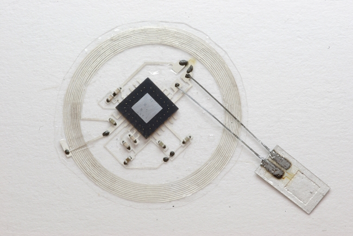
Wilson Ray and Washington University colleagues,  in partnership with John Rogers,  have developed a miniaturized wireless  device to monitor temperature and pressure when implanted into the brain following TBI.  The implant then dissolves, to be naturally reabsorbed into soft tissue, once  no longer needed.

Current methods involve an implanted sensor that must be hard-wired to an external monitoring instrument,  with risks of hemorrhage or infection, and requiring multiple rounds of surgery.

The technology has positive implications for various types of monitoring or therapeutic devices that are implanted or ingested.


Wearable Tech + Digital Health San Francisco – April 5, 2016 @ the Mission Bay Conference Center

NeuroTech San Francisco – April 6, 2016 @ the Mission Bay Conference Center

Wearable Tech + Digital Health NYC – June 7, 2016 @ the New York Academy of Sciences

NeuroTech NYC – June 8, 2016 @ the New York Academy of Sciences
 

Categories
Brain

Handheld spectrometer identifies low-grade brain tumors

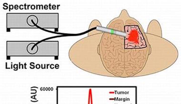
Emory and Georgia Tech researchers have developed a highly sensitive spectrometer  to identify low-grade gliomas from healthy tissue.  The hand-held device  contains a light source and detector tuned to the excitation and emission wavelengths of PpIX.

The team claims that the device is 3 times more sensitive than current surgical microscopes, and enables the detection of as few as 1000 tumor cells.

This could lead to the ability of surgeons to remove low-grade gliomas with fluorescence-guided procedures.


Wearable Tech + Digital Health San Francisco – April 5, 2016 @ the Mission Bay Conference Center

 NeuroTech San Francisco – April 6, 2016 @ the Mission Bay Conference Center

Wearable Tech + Digital Health NYC – June 7, 2016 @ the New York Academy of Sciences

NeuroTech NYC – June 8, 2016 @ the New York Academy of Sciences
 

Categories
Brain Wearables

Smart helmet + rate activated strap could minimize head injury severity

The Army Research Lab continues to develop technology aimed at protecting soldiers, athletes and others from impact related head injury.

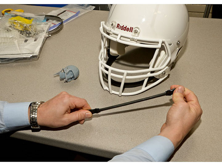
Its rate-activated helmet-strap can prevent violent head motions, while permitting (necessary) voluntary head motion. The material stretches with low, elastic force at slower speeds, and resists with high force when pulled quickly upon impact.

Used in combination with sensors that quantify impact in real time, and detect and monitor biological and physiological indicators of TBI, the strap system might be able to minimize injury.

The project is being funded by the NFL, GE, and  UnderArmour. Click to view the video of project manager Eric Wetzel describing the technology.


Wearable Tech + Digital Health San Francisco – April 5,2016 @ the Mission Bay Conference Center

NeuroTech San Francisco – April 6, 2016 @ the Misson Bay Conference Center

Wearable Tech + Digital Health NYC – June 7, 2016 @ the New York Academy of Sciences

NeuroTech NYC – June 8, 2016 @ the New York Academy of Sciences
 

Categories
AI Brain Deep Learning fMRI

Brain architecture linked to consciousness, abstract thought

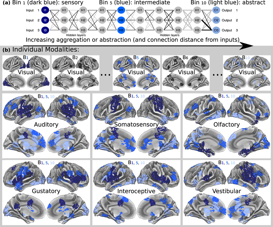
UMass professor Hava Siegelmann used fMRI data from tens of thousands of patients to understand how thought arises from brain structure. This resulted in a geometry-based  method meant to advance the identification and treatment of brain disease.  It can also be used to improve deep learning systems, and her lab is now creating a “massively recurrent deep learning network.”

Siegelmann found that cognitive function and abstract thought exist as an agglomeration of many cortical sources, from those close to sensory cortices to those far deeper along the brain connector. Her data-driven analyses defined a hierarchically ordered connectome, revealing a related continuum of cognitive function.

Siegelmann claims that  “with a slope (geometrical algorithm) identifier, behaviors could now be ordered by their relative depth activity with no human intervention or bias.”


Wearable Tech + Digital Health San Francisco – April 5, 2016 @ the Mission Bay Conference Center

NeuroTech San Francisco – April 6, 2016 @ the Mission Bay Conference Center

Wearable Tech + Digital Health NYC – June 7, 2016 @ the New York Academy of Sciences

NeuroTech NYC – June 8, 2016 @ the New York Academy of Sciences
 

Categories
Brain

CTE/sports head trauma link examined

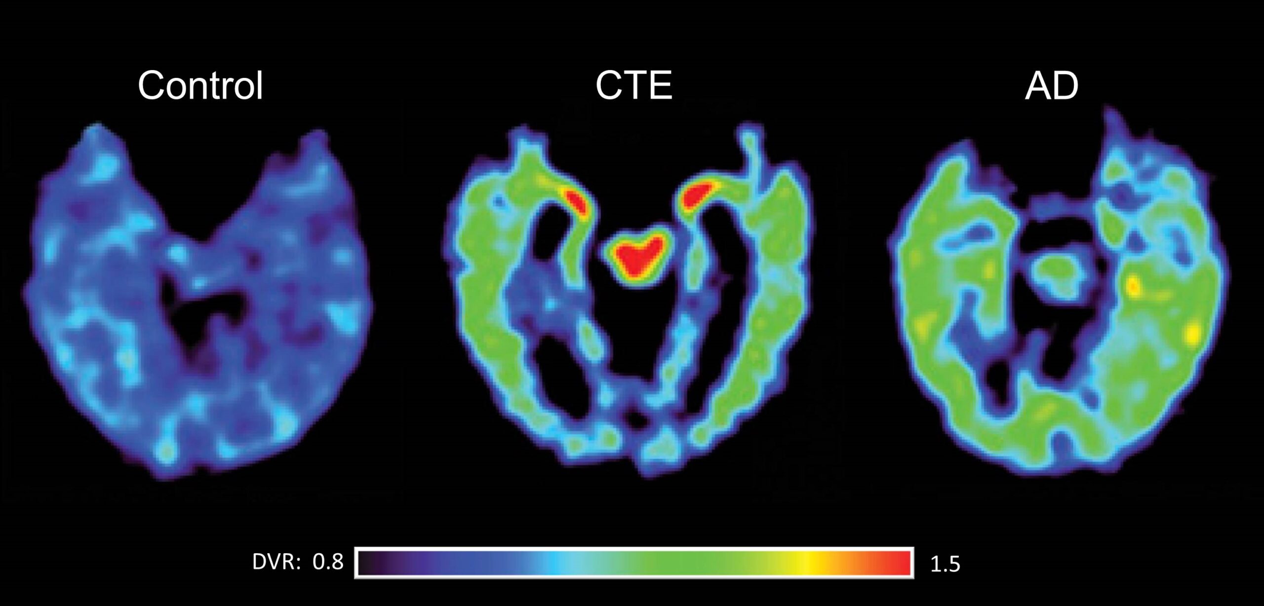
BU, Cleveland Clinic, Banner Alzheimer’s Institute and Brigham and Women’s have received a $16 million NIH grant to improve the detection and diagnosis of chronic traumatic encephalopathy, and examine risk factors for the disease.

The study is the first to examine CTE in living patients, with the goal of understanding the  link between CTE and head trauma.  The degenerative brain disease is linked to repeated head hits in contact sports. It is characterized by changes in behavior, mood and cognition, including the development of dementia. Currently it can only be diagnosed post-mortem.


Wearable Tech + Digital Health San Francisco – April 5, 2016 @ the Mission Bay Conference Center

NeuroTech San Francisco – April 6, 2016 @ the Mission Bay Conference Center

Wearable Tech + Digital Health NYC – June 7, 2016 @ the New York Academy of Sciences

NeuroTech NYC – June 8, 2016 @ the New York Academy of Sciences
Categories
Brain

Study: Thalamus stimulation alters activity, alertness

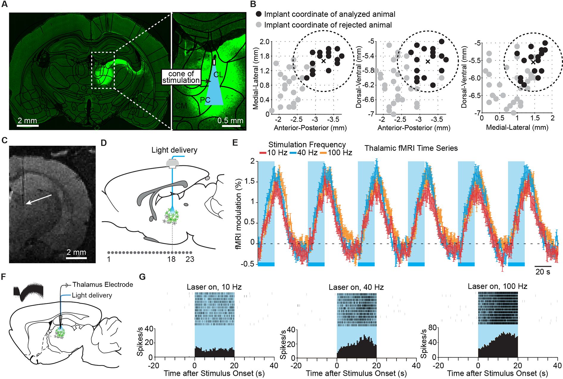
Stanford researchers have used optogenetic lasers targeted at the thalamus to turn whole-brain activity on and off in rats.  They hope that this will lead to improved optogenetic therapy for humans, as the thalamus controls memory, attention, and sleep.  The study was led by Jin Hyung Lee,  Hyun Joo Lee, Jia Liu, Andrew Weitz and Zhongnan Fang.

By flashing high-frequency optogenetic lasers at the thalamus, the team woke sleeping rats and cause widespread brain activity. When they flashed the laser at 10 pulses per second,  the activity of the brain’s sensory cortex was suppressed, and the and  rats entered a seizure-like state of unconsciousness.

A combination of optogenetics, fMRI, EEG and single-unit cell recordings  detected overall effects on the brain.

According to Lee,  “using targeted, temporally precise ontogenetic stimulation allowed us to selectively excite a single group of neuronal elements and identify their specific role in creating distinct modes of network function. ” This could not previously be achieved with conventional electrode stimulation.

The results suggest that the central thalamus can either power the brain to an “awake” state or promote a state of unconsciousness, depending on how rapidly its neurons are stimulated.


Wearable Tech + Digital Health San Francisco – April 5, 2016 @ the Mission Bay Conference Center

NeuroTech San Francisco – April 6, 2016 @ the Mission Bay Conference Center

Wearable Tech + Digital Health NYC – June 7, 2016 @ the New York Academy of Sciences

NeuroTech NYC – June 8, 2016 @ the New York Academy of Sciences
 

Categories
Brain Parkinson's Personalized Medicine Wearables

Implant + wearable to track neuromodulation effectiveness

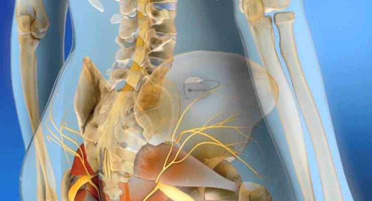
Medtronic is linking its implanted devices with Samsung’s phones and tablets to better monitor the effectiveness of neuromodulation technologies.  (Click to view Samsung release.)

Those with implanted neurostimulators, which  send electronic signals to targeted areas of the brain to block symptoms, can have a more active role in the management of their diseases.  Parkinson’s, essential tremor and dystonia patients will hopefully benefit from the initiative.

Data from the devices will be sent to a patient’s mobile devices, including phones, wearables and tablets, in real time.  It can also be sent directly to a doctor to help them better understand patient symptoms and progress, and appropriately adjust therapies.

The two companies announces a similar partnership for the management of diabetes earlier this year.


Wearable Tech + Digital Health San Francisco – April 5, 2016 @ the Mission Bay Conference Center

NeuroTech San Francisco – April 6, 2016 @ the Misson Bay Conference Center

Wearable Tech + Digital Health NYC – June 7, 2016 @ the New York Academy of Sciences
NeuroTech NYC – June 8, 2016 @ the New York Academy of Sciences