Categories
Brain

Military shockwave sensor used for sports concussion detection

Early detection of concussion can enable more rapid treatment and better outcomes.

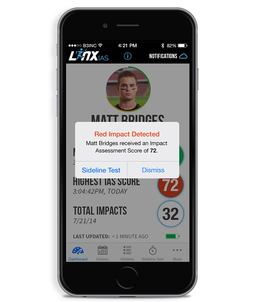
The Linx IAS  sensor detects head impact severity during sports training.  It was built upon military technology that measures the  impact of  explosion shockwaves on the brain.

The sensor is part of fabric headband.  Concussive forces are transmitted to an app that rates each blow from 1-99.  Each rating has an associated traffic light color code.  Green signals a low level “sub-concussive” blow, while red identifies a severe hit.

WEARABLE TECH + DIGITAL HEALTH SAN FRANCISCO – APRIL 5, 2016 @ THE MISSION BAY CONFERENCE CENTER

NEUROTECH SAN FRANCISCO – APRIL 6, 2016 @ THE MISSION BAY CONFERENCE CENTER
Categories
Autism Brain Wearables

Glass app helps autistic kids understand expressions, emotions

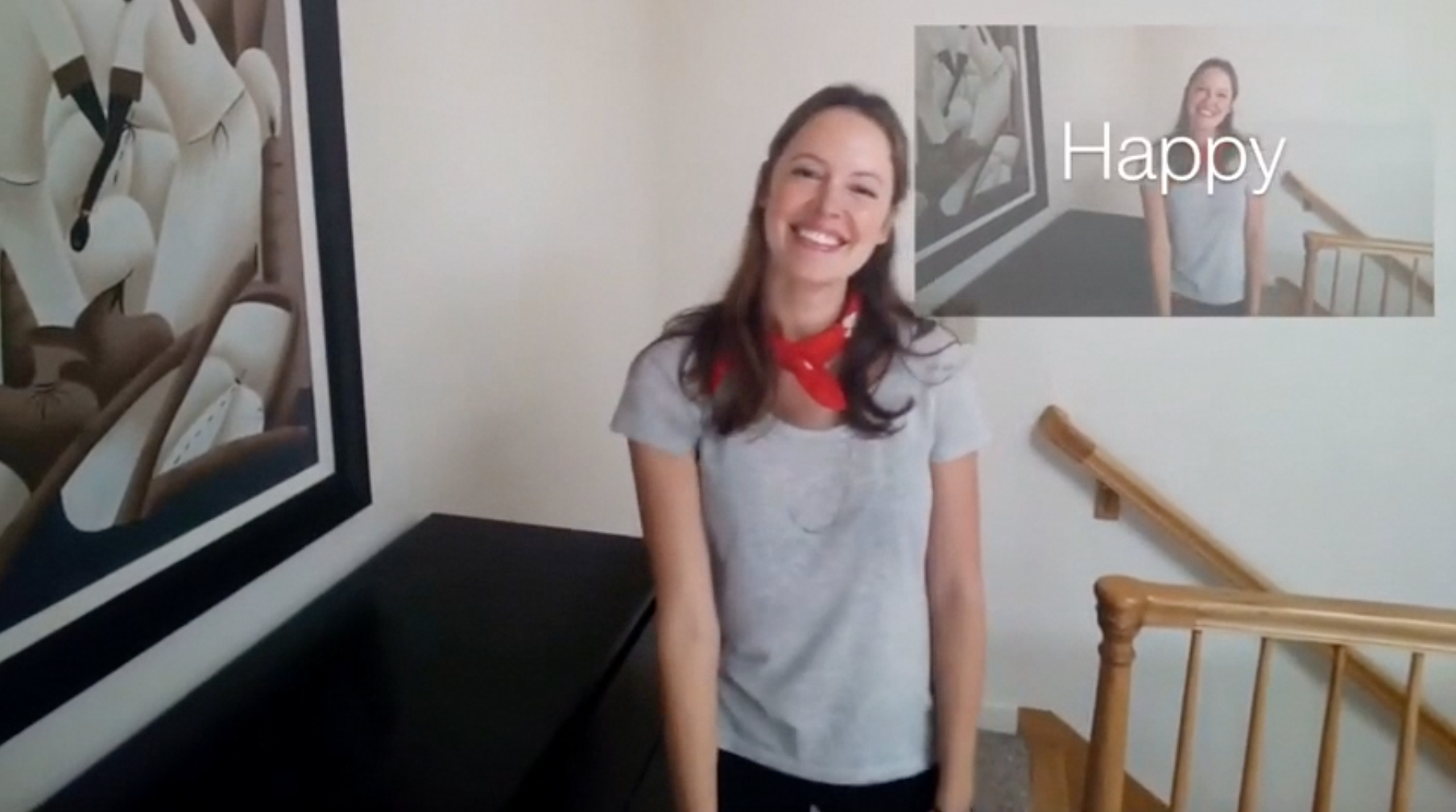
Dennis Wall, Catalin Voss, and  Nick Haber of Stanford’s Wall Lab are developing Google Glass software to help autistic children recognize and understand facial expressions and emotions.

Head motion tracking sensors, a microphone, and an eye tracking infrared camera analyze a wearer’s behavior during social interactions. Real time social cues are provided, and responses, including eye contact details, which can be analyzed in behavioral therapy, are recorded. The goal is to incorporate behavioral therapy into natural settings.

Last year ApplySci described a related technology,  Brain Power‘s Glass app, where expressions are interpreted and social engagement with parents is monitored, using games and exercises.

The Autism Glass Project has been tested on 40 children, and a clinical trial of 100 participants is now beginning.

WEARABLE TECH + DIGITAL HEALTH SAN FRANCISCO – APRIL 5, 2016 @ THE MISSION BAY CONFERENCE CENTER

NEUROTECH SAN FRANCISCO – APRIL 6, 2016 @ THE MISSION BAY CONFERENCE CENTER
Categories
Brain Parkinson's

Study: Cancer drug improves Parkinson’s cognitive, motor functions

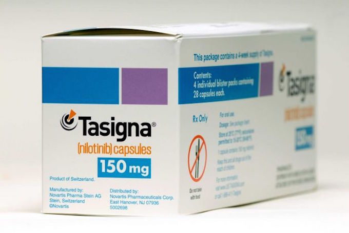
A small, early stage trial (with no control group) at Georgetown has  found that a small dose of the leukemia drug nilotinib (brand name “Tasigna” by Novartis) produced “meaningful clinical improvements” in 10 out of 11 patients.

The potential impact is significant, and the researchers believe that expanded studies will validate the  promising results. During the trial, participant dopamine levels increased so much that they were advised to reduce or stop taking other drugs.

The investigators reported that one participant, who was confined to a wheelchair,  was able to walk again, and three participants who could not speak were able to hold conversations.

The study marks the first time a therapy appears to reverse the “cognitive and motor decline in patients with these neuro-degenerative disorders,” according to Professor Fernando Pagan, who led the study with Charbel Moussa.

There has been some success with stimulation treatments for Parkison’s symptoms, and advances in early diagnosis and monitoring, but there is no known cure for this debilitating disease.  (See ApplySci Parkinson’s coverage, 2013-2015.)

Click to view Georgetown University Medical Center video.

WEARBLE TECH + DIGITAL HEALTH SAN FRANCISCO – APRIL 5, 2016 @ THE MISSION BAY CONFERENCE CENTER

NEUROTECH SAN FRANCISCO – APRIL 6, 2016 @ THE MISSION BAY CONFERENCE CENTER
Categories
Brain Prosthetics Sensors

Sensors allow more natural sense of touch in prosthetics

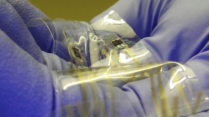
Stanford’s Zhenan Bao is developing technology that could restore a more natural sense of touch in prosthetics.  Her flexible, thin plastic sensors send signals to the brain that more closely resemble nerve messages of human skin touch sensors.

The disruptive technology has not yet been tested on humans, and researchers still need to find a safe way to pass electrical signals from prostheses to the brain for long periods.

Many teams are working toward this (see ApplySci coverage from 2013-2015).   Previous tactile sensors have however been analogue devices, where more pressure produces a stronger electrical signal, rather than a more frequent stream of pulses. The electrical signals must then be sent to another processing chip that converts the strength of the signals to a digital stream of pulses that is only then sent on to peripheral nerves or brain tissue.  Bao’s sensors send digital signals directly.

Click to view Stanford University video.

WEARABLE TECH + DIGITAL HEALTH SAN FRANCISCO – APRIL 5, 2016 @ THE MISSION BAY CONFERENCE CENTER

NEUROTECH SAN FRANCISCO – APRIL 6, 2016 @ THE MISSION BAY CONFERENCE CENTER
Categories
Brain

Bipolar mood detection via smartphone

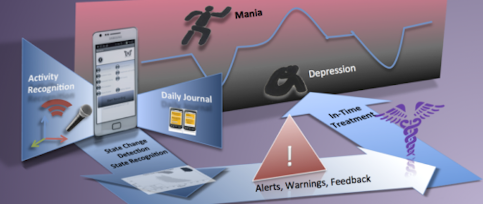
In July, ApplySci described a Northwestern developed phone app that monitors behavior patterns to detect depression.  Now, Venet Osmani at CREATE-NET has announced a similar phone based concept with a focus on bipolar disorder.   A small study has shown that mood changes can be accurately spotted as they occur,  facilitating earlier treatment and better outcomes.

The manic phase of the disease is often characterized by hyperactivity, which can be measured by an accelerometer, GPS device, speech analysis (for rapid speech) and phone records (for frequent conversations).

Patients in the depressive stage usually demonstrate distinctly different behaviors.

Smartphone activity of 12 bipolar patients was monitored over 12 weeks.  They visited the clinic every three weeks, when a conventional mental state evaluation occurred.

The study found that activity and location data gave a good indication of mood, and accurately predicted mood change 94 percent of the time. When combined with call and speech analysis, accuracy climbed to 97 per cent.  According to Osmani, “amost all changes were detected with almost no false alarms.”

WEARABLE TECH + DIGITAL HEALTH SAN FRANCISCO – APRIL 5, 2016 @ THE MISSION BAY CONFERENCE CENTER

NEUROTECH SAN FRANCISCO – APRIL 6, 2016 @ THE MISSION BAY CONFERENCE CENTER
Categories
Brain

Implant captures neural signals, leaves surrounding tissue intact

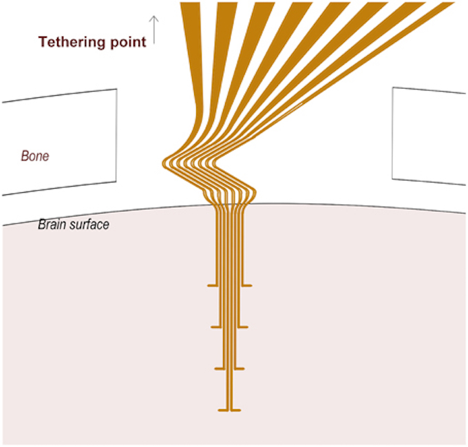
Lund professor Jens Schouenborg has developed implantable multichannel electrodes that can capture signals from single neurons over a long period — without causing brain tissue damage.  While not yet tested on humans, Schouenborg believes that the discovery will make it possible to understand brain function in both healthy and diseased individuals.  Potential applications include Parkinson’s and chronic pain treatments.

Current flexible electrodes cannnot maintain their shape when implanted, and must be attached to a solid chip, limiting their flexibility.  This irritates brain tissue, killing surrounding nerve cells and making signals unreliable.

NEUROTECH SAN FRANCISCO CONFERENCE – APRIL 6, 2016 @ MISSION BAY CONFERENCE CENTER

Categories
BCI Brain

DARPA peripheral nerve modulation project launches

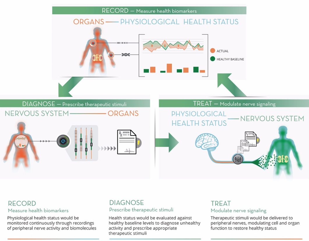
In September, 2014, ApplySci described DARPA’s proposed ElectRX (Electrical Prescriptions) project.  The agency has now selected 7 research teams  to begin work on the program, which is lead by Douglas Weber.  The goal is to develop a closed-loop system to treat disease by modulating the activity of peripheral nerves. The teams will work to develop a system, to be tested in human clinical trials, to treat chronic pain, inflammatory disease, post-traumatic stress and other illnesses.

The selected teams and their foci follow:

  • Circuit Therapeutics (Menlo Park), a start-up co-founded by Karl Deisseroth and Scott Delp, plans to further develop its experimental optogenetic methods for treating neuropathic pain, building toward testing in animal models before seeking to move to clinical trials in humans.
  • A team at Columbia University (New York), led by Elisa Konofagou, will pursue fundamental science to support the use of non-invasive, targeted ultrasound for neuromodulation. The team aims to elucidate the underlying mechanisms that may make ultrasound an option for chronic intervention, including activation and inhibition of nerves.
  • A team at the Florey Institute of Neuroscience and Mental Health (Australia), led by John Furness, will seek to map the nerve pathways that underlie intestinal inflammation, with a focus on determining the correlations between animal models and human neural circuitry. They will also explore the use of neurostimulation technologies based on the cochlear implant —developed by Cochlear, Inc. to treat hearing loss, but adapted to modulate activity of the vagus nerve in response to biofeedback signals—as a possible treatment for inflammatory bowel disease.
  • A team at the Johns Hopkins University (Baltimore), led by Jiande Chen, aims to explore the root mechanisms of inflammatory bowel disease and the impact of sacral nerve stimulation on its progression. The team will apply a first-of-its-kind approach to visualize intestinal responses to neuromodulation in animal models.
  • A team at the Massachusetts Institute of Technology (Cambridge), led by Polina Anikeeva, will aim to advance its established work in magnetic nanoparticles for localized, precision in vivo neuromodulation through thermal activation of neurons in animal models. The team’s work will target the adrenal gland and the splanchnic nerve circuits that govern its function. To increase specificity and minimize potential side effects of this method of stimulation, the team seeks to develop nanoparticles with the ability to bind to neuronal membranes.
  • A team at Purdue University (Indiana), led by Pedro Irazoqui, will leverage an existing collaboration with Cyberonics to study inflammation of the gastrointestinal tract and its responsiveness to vagal nerve stimulation through the neck. Validation of the mechanistic insights that emerge from the effort will take place in pre-clinical models in which novel neuromodulation devices will be applied to reduce inflammation in a feedback-controlled manner. Later stages of the effort could advance the design of clinical neuromodulation devices.
  • A team at the University of Texas, Dallas, led by Robert Rennaker and Michael Kilgard, will examine the use of vagal nerve stimulation to induce neural plasticity for the treatment of post-traumatic stress. As envisioned, stimulation could enhance learned behavioral responses that reduce fear and anxiety when presented with traumatic cues. Dr. Rennaker is a U.S. Marine Corps veteran who served in Liberia, Kuwait and Yugoslavia.

    NEUROTECH SAN FRANCISCO CONFERENCE – APRIL 6, 2016 @ THE MISSION BAY CONFERENCE CENTER
Categories
Brain

Cheap, accessible mini-brain for biomedical research

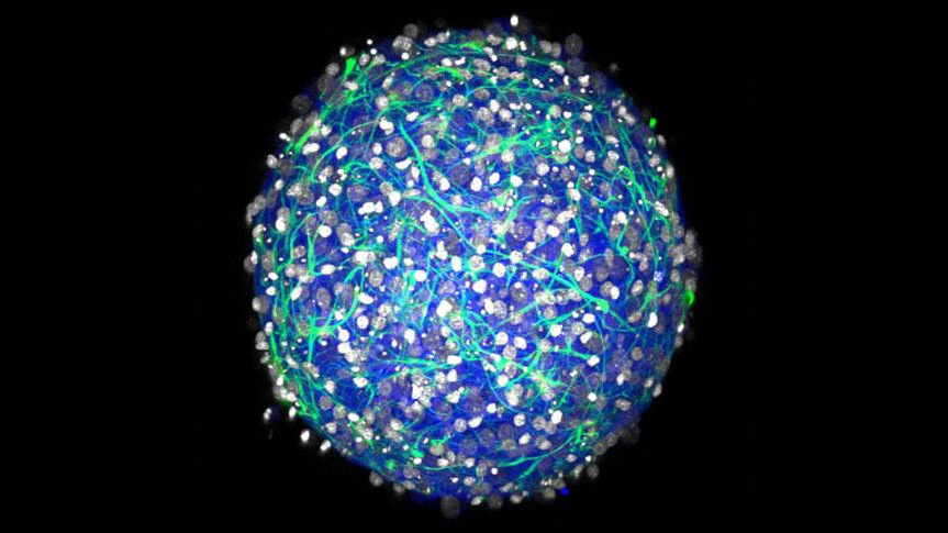
Brown University‘s Diane Hoffman-KimYu-Ting Dingle and Molly Boutin  have developed a cheap method for developing a 3D mini brain for biomedical research.

The central nervous system tissue sphere can produce electrical signals and form synapses.  Applications include drug testing,  neural tissue transplant testing, and stem cell experiments.

The mini-brains are not the first or most sophisticated working cell cultures of a central nervous system, but they require fewer steps to make and use readily available materials.  Dingle compares the technology to retail 3-D printers, which have proliferated, bringing once-rare technology to a mass market. “We could allow all kinds of labs to do this research,” she said.

The method yields mini-brains with several properties:

  • Diverse cell types: The cultures contain both inhibitory and excitatory neurons and several varieties of essential neural support cells called glia.
  • Electrically active: the neurons fire and spike and form synaptic connections, producing complex networks.
  • 3-D: Cells connect and communicate within a realistic geometry, rather than merely across a flat plane as in a 2-D culture.
  • Natural density: Experiments showed that the mini-brains have a density of a few hundred thousand cells per cubic millimeter, which is similar to a natural rodent brain.
  • Physical structure: Cells in the mini-brain produce their own extracellular matrix, producing a tissue with the same mechanical properties (squishiness) as natural tissue. The cultures also don’t rely on foreign materials such as scaffolds of collagen.
  • Longevity: In testing, cultured tissues live for at least a month.

The spheres of brain tissue begin to form within a day after the cultures are seeded, and  form complex 3-D neural networks in 2-3 weeks.

NEUROTECH SAN FRANCISCO CONFERENCE – APRIL 6, 2016 – EARLY REGISTRATION ENDS OCTOBER 17TH.

Categories
BCI Brain

Brain-to-brain link allows one person to read another’s thoughts

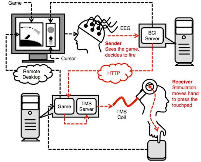
University of Washington researchers used a direct brain-to-brain connection to enable pairs of participants to play a question-and-answer game by transmitting signals from one brain to the other over the Internet. The experiment is thought to be the first to show that two brains can be directly linked to allow one person to guess what’s on another person’s mind.

Lead author Andrea Stocco believes  that “This is the most complex brain-to-brain experiment that’s been done to date in humans. It uses conscious experiences through signals that are experienced visually, and it requires two people to collaborate.”  Chanel Prat, Darbey Losey, Jeneva Cronin, Joseph Wu and Justin Abernathy co-authored the paper.

The study builds on the UW team’s 2013 experiment demonstrating a direct brain-to-brain connection between humans. Other scientists have connected the brains of rats and monkeys, and transmitted brain signals from a human to a rat, using electrodes inserted into animals’ brains. The UW team used noninvasive technology to send a person’s brain signals over the Internet to control the hand motions of another person.

Click to watch the University of Washington video.

Categories
Assistive Technologies Brain Gaming

Virtual coaching for TBI patients

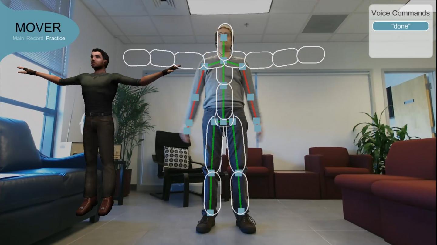
The Office of Naval Research is developing MOVER (Mobile, Virtual Enhancements for Rehabilitation) to help TBI patients maintain therapy regimens.  Confusion, forgetfulness or depression can prevent injured veterans from completing necessary exercises for rehabilitation. Featured movements include including lunges, knee raises and squats, which are standard for TBI therapy.

When a user turns on a computer and camera, he/she stands still, while MOVER maps a virtual “skeleton” of brightly colored lines and shapes.  Movements are mirrored through each exercise. To increase visibility, users can connect MOVER to a television using Microsoft Kinect.

The system coaches by displaying pop-up text boxes or color shading in areas of the virtual skeleton, highlighting where and how to correct one’s form.

A six-month pilot study of the software, with 40 TBI patients and therapists at Spaulding Rehabilitation Hospital, will soon begin.

Categories
Brain Ultrasound

Sonogenetics: Neuron stimulation via ultrasound

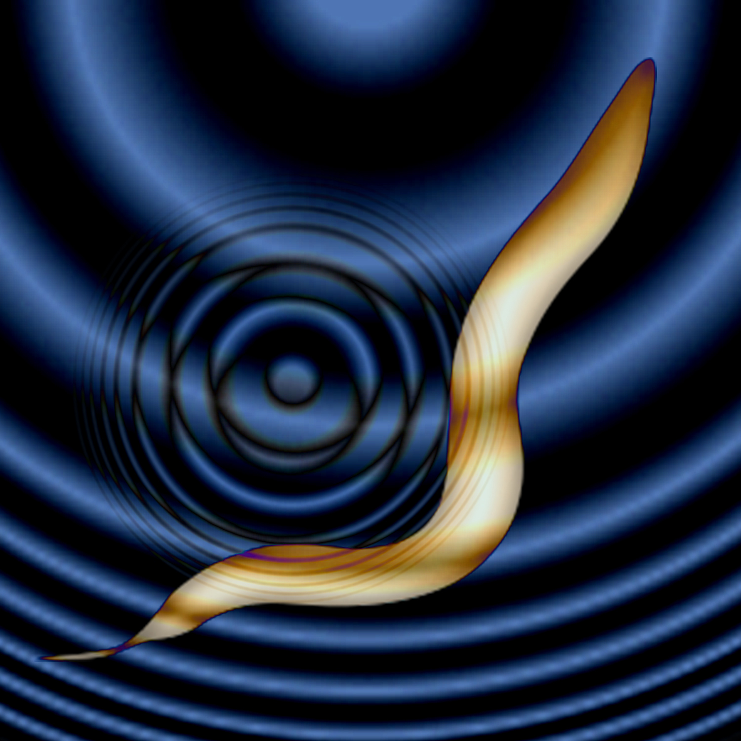
Salk‘s Sreekanth Chalasani‘s “sonogenetics” technique uses ultrasound to stimulate individual brain cells.  A nature paper describes the technology as tested on worms.  The goal is noninvasive stimulation of specific cell types or individual neurons in humans, with out using implanted electrodes or fiber-optic cables.

Current optogenetics therapies  rely on inserting light-sensitive channel proteins into neurons. When hit by the correct color of light, usually sent by a fiber-optic cable, the channels open, allowing ions to flood in.

The new technique relies on touch-sensitive  “channel” proteins, which can be added to specific brain cells through genetic engineering. The channels open when hit by an ultrasonic pulse, allowing ions to flood into a neuron and cause it to turn on.

Click to view Salk Institute video.

Categories
Apps Brain Gaming

Sport performance game app to train the brain

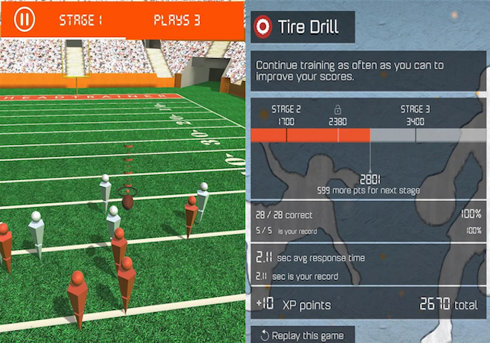
Games are increasingly recognized as a method of enhancing cognitive abilities.  HeadTrainer is meant to improve the brain with 5-10 minutes of daily gameplay.

The sports games were designed to exercise 5 cognitive skills:   decision making, processing speed, focus, memory, and visual/spatial awareness.  Developer Deborah Attix of Duke University focused on testing and training the brain with each game.

The company believes that in the future, HeadTrainer and similar apps can contribute to the mental, emotional, health, and hormonal metrics that are key to sports analytics.