Categories
Brain Parkinson's

Study: Cancer drug improves Parkinson’s cognitive, motor functions

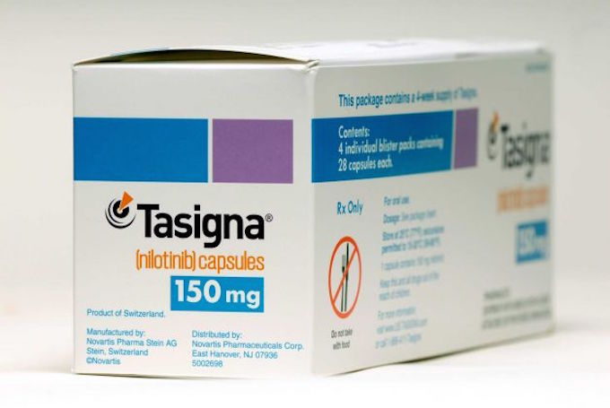
A small, early stage trial (with no control group) at Georgetown has  found that a small dose of the leukemia drug nilotinib (brand name “Tasigna” by Novartis) produced “meaningful clinical improvements” in 10 out of 11 patients.

The potential impact is significant, and the researchers believe that expanded studies will validate the  promising results. During the trial, participant dopamine levels increased so much that they were advised to reduce or stop taking other drugs.

The investigators reported that one participant, who was confined to a wheelchair,  was able to walk again, and three participants who could not speak were able to hold conversations.

The study marks the first time a therapy appears to reverse the “cognitive and motor decline in patients with these neuro-degenerative disorders,” according to Professor Fernando Pagan, who led the study with Charbel Moussa.

There has been some success with stimulation treatments for Parkison’s symptoms, and advances in early diagnosis and monitoring, but there is no known cure for this debilitating disease.  (See ApplySci Parkinson’s coverage, 2013-2015.)

Click to view Georgetown University Medical Center video.

WEARBLE TECH + DIGITAL HEALTH SAN FRANCISCO – APRIL 5, 2016 @ THE MISSION BAY CONFERENCE CENTER

NEUROTECH SAN FRANCISCO – APRIL 6, 2016 @ THE MISSION BAY CONFERENCE CENTER
Categories
Brain Prosthetics Sensors

Sensors allow more natural sense of touch in prosthetics

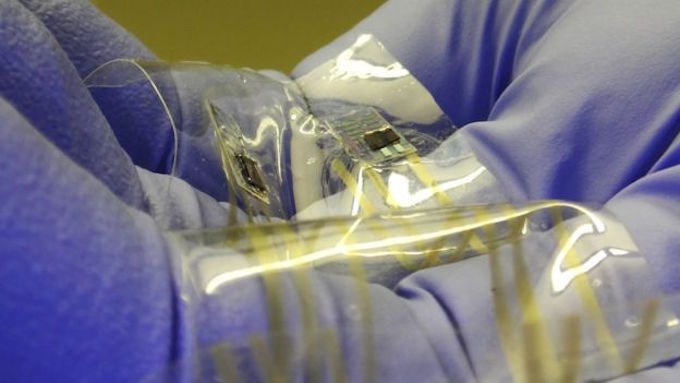
Stanford’s Zhenan Bao is developing technology that could restore a more natural sense of touch in prosthetics.  Her flexible, thin plastic sensors send signals to the brain that more closely resemble nerve messages of human skin touch sensors.

The disruptive technology has not yet been tested on humans, and researchers still need to find a safe way to pass electrical signals from prostheses to the brain for long periods.

Many teams are working toward this (see ApplySci coverage from 2013-2015).   Previous tactile sensors have however been analogue devices, where more pressure produces a stronger electrical signal, rather than a more frequent stream of pulses. The electrical signals must then be sent to another processing chip that converts the strength of the signals to a digital stream of pulses that is only then sent on to peripheral nerves or brain tissue.  Bao’s sensors send digital signals directly.

Click to view Stanford University video.

WEARABLE TECH + DIGITAL HEALTH SAN FRANCISCO – APRIL 5, 2016 @ THE MISSION BAY CONFERENCE CENTER

NEUROTECH SAN FRANCISCO – APRIL 6, 2016 @ THE MISSION BAY CONFERENCE CENTER
Categories
Epilepsy Wearables

App detects seizure onset with heart rate, accelerometer data

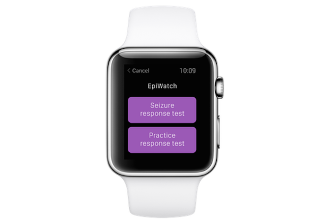
Johns Hopkins professor Gregory Krauss has used ResearchKit to develop an app to detect the onset and duration of epileptic seizures with an Apple Watch.  Wearers must touch the watch to capture accelerometer and heart rate sensor data, and notify a caregiver.  The EpiWatch app logs seizures and responses, and tracks medication adherence and side effects.

The EpiWatch is similar in function to Embrace by Empatica, developed by MIT professor Rosalind Picard.  Embrace uses skin conductance, accelerator, and gyrometer  data to detect seizures.  (See ApplySci, November 28, 2014.)

ApplySci applauds these advances, which by recording patterns and notifying loved ones,  have the potential to improve the lives of epilepsy sufferers.  There is little evidence that cardiac activity alone can be used to predict seizures (see Amir Geva‘s paper in IEEE Transactions on Biomedical Engineering).  The next step is to develop the ability to predict seizures in advance, which will require brain activity interpretation.

WEARABLE TECH + DIGITAL HEALTH SAN FRANCISCO – APRIL 5, 2016 @ MISSION BAY CONFERENCE CENTER

NEUROTECH SAN FRANCISCO – APRIL 6, 2016 @ MISSION BAY CONFERENCE CENTER

Categories
fitness Smart Fabric

Sensor shorts provide real time runner feedback

ApplySci has described several examples of smart shirts and smart fabrics in recent months.  Now, Lumo his integrated sensors into shorts to monitor  cadence, stride length, pace, distance and pelvic rotation in runners.  Placed inside the waistband, the sensors sync with smartphones to provide real time feedback, and the app sends data and coaching content post-run.

WEARABLE TECH + DIGITAL HEALTH SAN FRANCISCO – APRIL 5, 2016 @ MISSION BAY CONFERENCE CENTER

NEUROTECH SAN FRANCISCO – APRIL 6, 2016 @ MISSION BAY CONFERENCE CENTER

Categories
Brain

Bipolar mood detection via smartphone

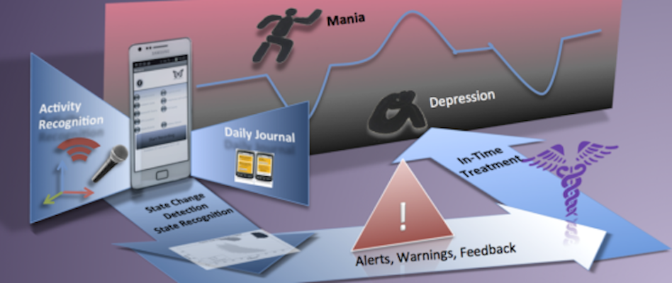
In July, ApplySci described a Northwestern developed phone app that monitors behavior patterns to detect depression.  Now, Venet Osmani at CREATE-NET has announced a similar phone based concept with a focus on bipolar disorder.   A small study has shown that mood changes can be accurately spotted as they occur,  facilitating earlier treatment and better outcomes.

The manic phase of the disease is often characterized by hyperactivity, which can be measured by an accelerometer, GPS device, speech analysis (for rapid speech) and phone records (for frequent conversations).

Patients in the depressive stage usually demonstrate distinctly different behaviors.

Smartphone activity of 12 bipolar patients was monitored over 12 weeks.  They visited the clinic every three weeks, when a conventional mental state evaluation occurred.

The study found that activity and location data gave a good indication of mood, and accurately predicted mood change 94 percent of the time. When combined with call and speech analysis, accuracy climbed to 97 per cent.  According to Osmani, “amost all changes were detected with almost no false alarms.”

WEARABLE TECH + DIGITAL HEALTH SAN FRANCISCO – APRIL 5, 2016 @ THE MISSION BAY CONFERENCE CENTER

NEUROTECH SAN FRANCISCO – APRIL 6, 2016 @ THE MISSION BAY CONFERENCE CENTER

Categories
Babies

Smart bottle monitors infant swallowing

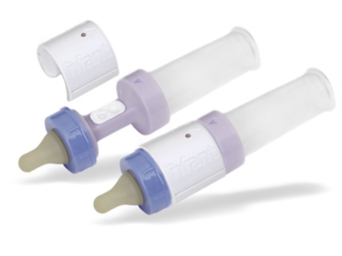
A smart baby bottle by nfant uses sensors to measure a baby’s tongue strength while feeding.  Data is sent to a caregiver’s phone and stored in the cloud.  Tongue movements determine whether a baby in the NICU has the strength to switch from tube to bottle or breastfeeding.

ApplySci sees the  opportunity for the next generation of smart bottles to incorporate multiple infant-health parameters, such as those derived from saliva.

WEARABLE TECH + DIGITAL HEALTH SAN FRANCISCO – APRIL 5, 2016 @ MISSION BAY CONFERENCE CENTER

NEUROTECH SAN FRANCISCO – APRIL 6, 2016 @ MISSION BAY CONFERENCE CENTER

Categories
Brain

Implant captures neural signals, leaves surrounding tissue intact

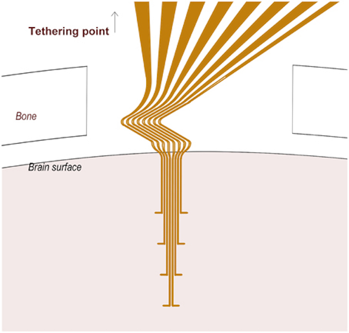
Lund professor Jens Schouenborg has developed implantable multichannel electrodes that can capture signals from single neurons over a long period — without causing brain tissue damage.  While not yet tested on humans, Schouenborg believes that the discovery will make it possible to understand brain function in both healthy and diseased individuals.  Potential applications include Parkinson’s and chronic pain treatments.

Current flexible electrodes cannnot maintain their shape when implanted, and must be attached to a solid chip, limiting their flexibility.  This irritates brain tissue, killing surrounding nerve cells and making signals unreliable.

NEUROTECH SAN FRANCISCO CONFERENCE – APRIL 6, 2016 @ MISSION BAY CONFERENCE CENTER

Categories
Heart

Noninvasive 3D scan identifies coronary artery blockages

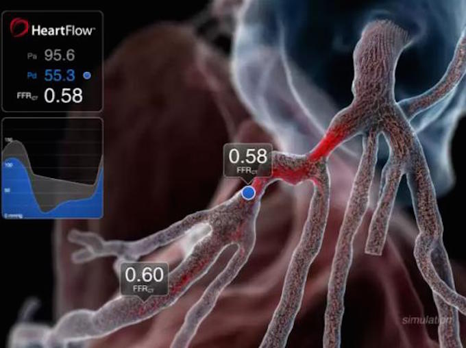
Fractional flow reserve computed tomography is a high-definition 3D scanner used to identify blocked arteries around the heart.  The technology, developed by Heartflow, eliminates the risk of diagnostic methods where vessels are probed.

The risk of death from an angiogram is one in 1,000, and can be caused by a rupture of an artery, or a stroke or heart attack caused by fatty plaque breaking off during the test.

FFRCT creates a detailed computer model of the heart from a CT image. It then (noninvasively) calculates the extent of blockages in the coronary arteries and whether they are restricting the flow of blood.

WEARABLE TECH + DIGITAL HEALTH SAN FRANCISCO – 4/5/16 @ MISSION BAY CONFERENCE CENTER

NEUROTECH SAN FRANCISCO – 4/6/16 @ MISSION BAY CONFERENCE CENTER

Categories
IoT Wearables

Swallowed sensors interact with smart devices

At a recent conference, Jawbone CEO Hosain Rahman said that his company is researching swallowable and implantable fitness sensors.  They would remain in a user’s bloodstream and be capable of monitoring multiple factors. The sensors could interact with smart devices, including adjusting thermostats if one’s body is too warm or cold, or not turning a car on if one’s blood/alcohol level is too high. While ApplySci believes that invasive, implanted sensors are not necessary for fitness/lifestyle applications, we applaud Jawbone’s work toward innovative, noninvasive devices

WEARABLE TECH + DIGITAL HEALTH SAN FRANCISCO – 4/5/16 @ MISSION BAY CONFERENCE CENTER

NEUROTECH SAN FRANCISCO – 4/6/16 @ MISSION BAY CONFERENCE CENTER

Categories
BCI Brain

DARPA peripheral nerve modulation project launches

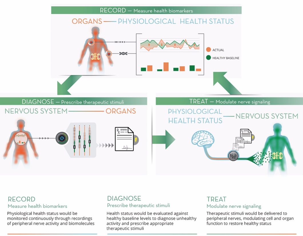
In September, 2014, ApplySci described DARPA’s proposed ElectRX (Electrical Prescriptions) project.  The agency has now selected 7 research teams  to begin work on the program, which is lead by Douglas Weber.  The goal is to develop a closed-loop system to treat disease by modulating the activity of peripheral nerves. The teams will work to develop a system, to be tested in human clinical trials, to treat chronic pain, inflammatory disease, post-traumatic stress and other illnesses.

The selected teams and their foci follow:

  • Circuit Therapeutics (Menlo Park), a start-up co-founded by Karl Deisseroth and Scott Delp, plans to further develop its experimental optogenetic methods for treating neuropathic pain, building toward testing in animal models before seeking to move to clinical trials in humans.
  • A team at Columbia University (New York), led by Elisa Konofagou, will pursue fundamental science to support the use of non-invasive, targeted ultrasound for neuromodulation. The team aims to elucidate the underlying mechanisms that may make ultrasound an option for chronic intervention, including activation and inhibition of nerves.
  • A team at the Florey Institute of Neuroscience and Mental Health (Australia), led by John Furness, will seek to map the nerve pathways that underlie intestinal inflammation, with a focus on determining the correlations between animal models and human neural circuitry. They will also explore the use of neurostimulation technologies based on the cochlear implant —developed by Cochlear, Inc. to treat hearing loss, but adapted to modulate activity of the vagus nerve in response to biofeedback signals—as a possible treatment for inflammatory bowel disease.
  • A team at the Johns Hopkins University (Baltimore), led by Jiande Chen, aims to explore the root mechanisms of inflammatory bowel disease and the impact of sacral nerve stimulation on its progression. The team will apply a first-of-its-kind approach to visualize intestinal responses to neuromodulation in animal models.
  • A team at the Massachusetts Institute of Technology (Cambridge), led by Polina Anikeeva, will aim to advance its established work in magnetic nanoparticles for localized, precision in vivo neuromodulation through thermal activation of neurons in animal models. The team’s work will target the adrenal gland and the splanchnic nerve circuits that govern its function. To increase specificity and minimize potential side effects of this method of stimulation, the team seeks to develop nanoparticles with the ability to bind to neuronal membranes.
  • A team at Purdue University (Indiana), led by Pedro Irazoqui, will leverage an existing collaboration with Cyberonics to study inflammation of the gastrointestinal tract and its responsiveness to vagal nerve stimulation through the neck. Validation of the mechanistic insights that emerge from the effort will take place in pre-clinical models in which novel neuromodulation devices will be applied to reduce inflammation in a feedback-controlled manner. Later stages of the effort could advance the design of clinical neuromodulation devices.
  • A team at the University of Texas, Dallas, led by Robert Rennaker and Michael Kilgard, will examine the use of vagal nerve stimulation to induce neural plasticity for the treatment of post-traumatic stress. As envisioned, stimulation could enhance learned behavioral responses that reduce fear and anxiety when presented with traumatic cues. Dr. Rennaker is a U.S. Marine Corps veteran who served in Liberia, Kuwait and Yugoslavia.

    NEUROTECH SAN FRANCISCO CONFERENCE – APRIL 6, 2016 @ THE MISSION BAY CONFERENCE CENTER
Categories
Heart

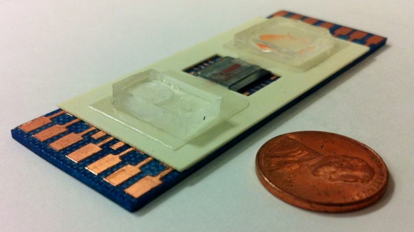
Device detects heart attacks with one drop of blood

UCLA’s Chi On Chui has developed a device that brings lab quality bio-molecular assessments to point-of-care settings, such as clinics, ambulances or homes.

SELFA (Semiconductor Electronic Label-Free Assay) could reduce emergency room time for heart attack patients by hours. Similar to a diabetic glucose sensor, SELFA uses a single drop of blood, taken wherever symptoms occur, to determine which patients do not require further medical care.

According to Chui: “Eighty-five percent of patients who present with symptoms of acute coronary syndrome in emergency rooms across the U.S. and Europe — some 12.75 million per year — have actually not had heart attacks after a standard test comes back, hours later, from the lab.”

WEARABLE TECH + DIGITAL HEALTH SAN FRANCISCO – APRIL 5, 2016 – REGISTER HERE

NEUROTECH SAN FRANCISCO – APRIL 6, 2016 – REGISTER HERE

Categories
Brain

Cheap, accessible mini-brain for biomedical research

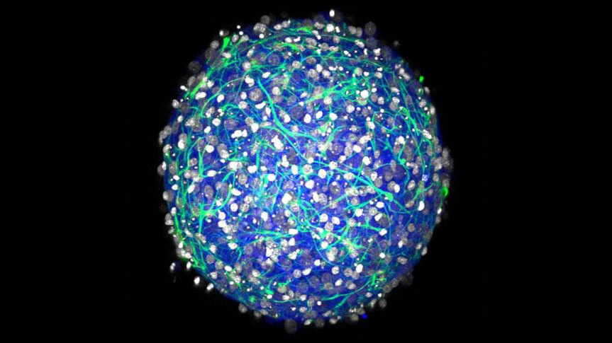
Brown University‘s Diane Hoffman-Kim, Yu-Ting Dingle and Molly Boutin  have developed a cheap method for developing a 3D mini brain for biomedical research.

The central nervous system tissue sphere can produce electrical signals and form synapses.  Applications include drug testing,  neural tissue transplant testing, and stem cell experiments.

The mini-brains are not the first or most sophisticated working cell cultures of a central nervous system, but they require fewer steps to make and use readily available materials.  Dingle compares the technology to retail 3-D printers, which have proliferated, bringing once-rare technology to a mass market. “We could allow all kinds of labs to do this research,” she said.

The method yields mini-brains with several properties:

  • Diverse cell types: The cultures contain both inhibitory and excitatory neurons and several varieties of essential neural support cells called glia.
  • Electrically active: the neurons fire and spike and form synaptic connections, producing complex networks.
  • 3-D: Cells connect and communicate within a realistic geometry, rather than merely across a flat plane as in a 2-D culture.
  • Natural density: Experiments showed that the mini-brains have a density of a few hundred thousand cells per cubic millimeter, which is similar to a natural rodent brain.
  • Physical structure: Cells in the mini-brain produce their own extracellular matrix, producing a tissue with the same mechanical properties (squishiness) as natural tissue. The cultures also don’t rely on foreign materials such as scaffolds of collagen.
  • Longevity: In testing, cultured tissues live for at least a month.

The spheres of brain tissue begin to form within a day after the cultures are seeded, and  form complex 3-D neural networks in 2-3 weeks.

NEUROTECH SAN FRANCISCO CONFERENCE – APRIL 6, 2016 – EARLY REGISTRATION ENDS OCTOBER 17TH.